Saloni Desai, Jyotirmay Biswas, Sudha K Ganesh, Darshan Bhatt
{"title":"Anterior segment optical coherence tomography: A monitoring tool in anterior scleritis.","authors":"Saloni Desai, Jyotirmay Biswas, Sudha K Ganesh, Darshan Bhatt","doi":"10.4103/ojo.ojo_376_24","DOIUrl":null,"url":null,"abstract":"<p><strong>Purpose: </strong>The purpose of this study was to evaluate the role of anterior segment optical coherence tomography (ASOCT) in the diagnosis and management of anterior scleritis.</p><p><strong>Patients and methods: </strong>In this retrospective study, we examined 58 eyes of 44 patients with anterior scleritis. The unaffected eye served as a control. In bilateral cases, the less affected eye was taken as the control. ASOCT image over the inflamed area and over the corresponding same area in the control eye was taken. The images were analysed for the presence or absence of hyporeflective areas, and the mean total scleral thickness (MTST) was measured. Both the images were then compared.</p><p><strong>Results: </strong>The mean age of our cohort was 51 ± 14.57 years. There were 14 males and 30 females. 68.18% (<i>n</i> = 30) were unilateral cases. The mean duration of anterior scleritis was 55.3 months, with 50% (<i>n</i> = 22) of patients having diffuse anterior scleritis. The majority of patients were treated with oral steroids (97.7%, <i>n</i> = 43) with or without the combination of immunosuppressant and biologics. The MTST during active disease (922.17 μm ± 252.03 μm) was statistically higher than the control group (798.05 μm ± 150.61 μm) (<i>P</i> = 0.005). The MTST in unilateral cases during active disease was 929.88 μm, which was significantly higher than in the control eyes (801.65 μm) (<i>P</i> = 0.02). There were 31 recurrent cases, of which 41.9% (<i>n</i> = 13) showed scleral thinning, and the mean scleral thinning in recurrent cases after treatment was 86.71 μm.</p><p><strong>Conclusion: </strong>ASOCT serves as a useful qualitative and quantitative tool for monitoring of patients with anterior scleritis under treatment.</p>","PeriodicalId":19461,"journal":{"name":"Oman Journal of Ophthalmology","volume":"18 2","pages":"155-161"},"PeriodicalIF":0.0000,"publicationDate":"2025-06-24","publicationTypes":"Journal Article","fieldsOfStudy":null,"isOpenAccess":false,"openAccessPdf":"https://www.ncbi.nlm.nih.gov/pmc/articles/PMC12258829/pdf/","citationCount":"0","resultStr":null,"platform":"Semanticscholar","paperid":null,"PeriodicalName":"Oman Journal of Ophthalmology","FirstCategoryId":"1085","ListUrlMain":"https://doi.org/10.4103/ojo.ojo_376_24","RegionNum":0,"RegionCategory":null,"ArticlePicture":[],"TitleCN":null,"AbstractTextCN":null,"PMCID":null,"EPubDate":"2025/5/1 0:00:00","PubModel":"eCollection","JCR":"Q3","JCRName":"Medicine","Score":null,"Total":0}
引用次数: 0
Abstract
Purpose: The purpose of this study was to evaluate the role of anterior segment optical coherence tomography (ASOCT) in the diagnosis and management of anterior scleritis.
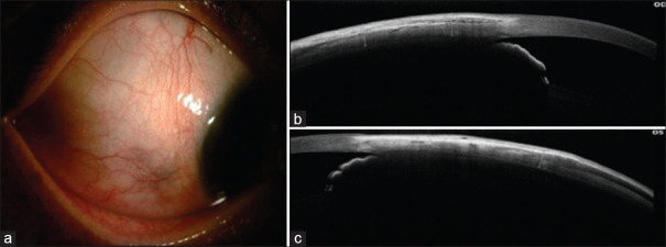
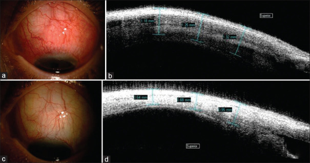
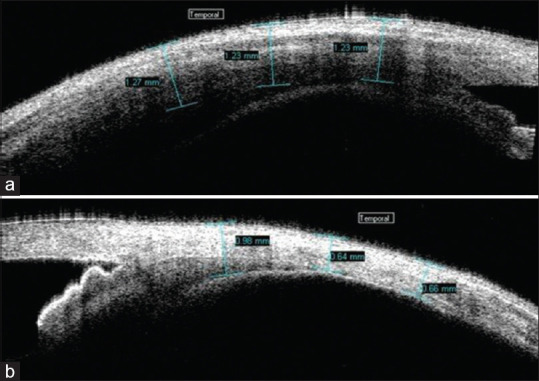
Patients and methods: In this retrospective study, we examined 58 eyes of 44 patients with anterior scleritis. The unaffected eye served as a control. In bilateral cases, the less affected eye was taken as the control. ASOCT image over the inflamed area and over the corresponding same area in the control eye was taken. The images were analysed for the presence or absence of hyporeflective areas, and the mean total scleral thickness (MTST) was measured. Both the images were then compared.
Results: The mean age of our cohort was 51 ± 14.57 years. There were 14 males and 30 females. 68.18% (n = 30) were unilateral cases. The mean duration of anterior scleritis was 55.3 months, with 50% (n = 22) of patients having diffuse anterior scleritis. The majority of patients were treated with oral steroids (97.7%, n = 43) with or without the combination of immunosuppressant and biologics. The MTST during active disease (922.17 μm ± 252.03 μm) was statistically higher than the control group (798.05 μm ± 150.61 μm) (P = 0.005). The MTST in unilateral cases during active disease was 929.88 μm, which was significantly higher than in the control eyes (801.65 μm) (P = 0.02). There were 31 recurrent cases, of which 41.9% (n = 13) showed scleral thinning, and the mean scleral thinning in recurrent cases after treatment was 86.71 μm.
Conclusion: ASOCT serves as a useful qualitative and quantitative tool for monitoring of patients with anterior scleritis under treatment.
期刊介绍:
To provide a platform for scientific expression of the Oman Ophthalmic Society and the international Ophthalmic community and to provide opportunities for free exchange of ideas and information. To serve as a valuable resource for ophthalmologists, eye-care providers including optometrists, orthoptists, other health care professionals and research workers in all aspects of the field of visual science.

 求助内容:
求助内容: 应助结果提醒方式:


