Vignesh J Krishnan, Sam Karimaghaei, Sami H Uwaydat
{"title":"A Case of Epiretinal Membrane Masquerading as Foveal Hypoplasia Uncovered by Optical Coherence Tomography Angiography.","authors":"Vignesh J Krishnan, Sam Karimaghaei, Sami H Uwaydat","doi":"10.1159/000546622","DOIUrl":null,"url":null,"abstract":"<p><strong>Introduction: </strong>This case report describes a patient whose misdiagnosis of foveal hypoplasia was uncovered by optical coherence tomography angiography (OCTA) findings that suggested the presence of an epiretinal membrane (ERM) over foveal hypoplasia.</p><p><strong>Case presentation: </strong>A 67-year-old man with no significant past medical history was referred to our retina clinic with a diagnosis of foveal hypoplasia. He had been experiencing significant vision loss for more than 1 year. OCT demonstrated absence of the foveal depression in both eyes. A subtle ERM was identified in the left eye OCT, but the presence of an ERM in the right eye OCT was equivocal. As such, it was unclear whether flattening of the fovea was attributable to hypoplasia or ERM based on OCT alone. This prompted further investigation with OCTA, which showed the presence of the FAZ in both eyes. The diagnosis of stage 2 ERM OU was made based on OCTA findings. The patient underwent cataract extraction with intraocular lens implantation, pars plana vitrectomy, and ERM peel, which resulted in improvement of visual symptoms and visual acuity. Follow-up OCT showed normalization of the foveal pit in the right eye greater than the left eye.</p><p><strong>Conclusion: </strong>This case demonstrates the importance of utilization of OCTA in differentiating true foveal hypoplasia from this foveal \"pseudo-hypoplasia\" exhibited by our patient.</p>","PeriodicalId":9635,"journal":{"name":"Case Reports in Ophthalmology","volume":"16 1","pages":"503-509"},"PeriodicalIF":0.6000,"publicationDate":"2025-06-23","publicationTypes":"Journal Article","fieldsOfStudy":null,"isOpenAccess":false,"openAccessPdf":"https://www.ncbi.nlm.nih.gov/pmc/articles/PMC12263143/pdf/","citationCount":"0","resultStr":null,"platform":"Semanticscholar","paperid":null,"PeriodicalName":"Case Reports in Ophthalmology","FirstCategoryId":"1085","ListUrlMain":"https://doi.org/10.1159/000546622","RegionNum":0,"RegionCategory":null,"ArticlePicture":[],"TitleCN":null,"AbstractTextCN":null,"PMCID":null,"EPubDate":"2025/1/1 0:00:00","PubModel":"eCollection","JCR":"Q4","JCRName":"OPHTHALMOLOGY","Score":null,"Total":0}
引用次数: 0
Abstract
Introduction: This case report describes a patient whose misdiagnosis of foveal hypoplasia was uncovered by optical coherence tomography angiography (OCTA) findings that suggested the presence of an epiretinal membrane (ERM) over foveal hypoplasia.
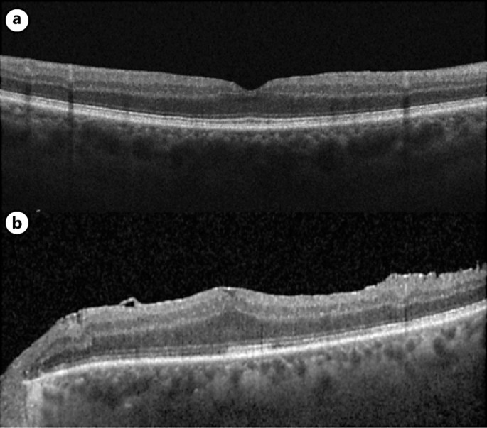
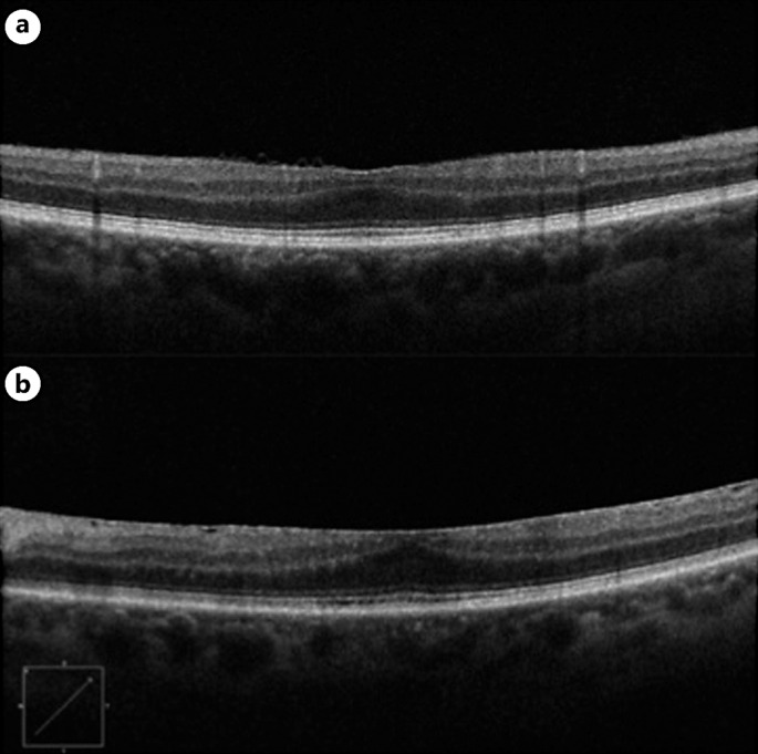
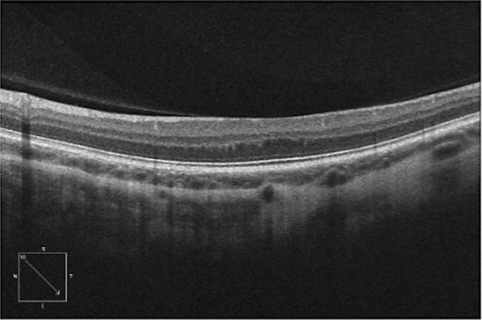
Case presentation: A 67-year-old man with no significant past medical history was referred to our retina clinic with a diagnosis of foveal hypoplasia. He had been experiencing significant vision loss for more than 1 year. OCT demonstrated absence of the foveal depression in both eyes. A subtle ERM was identified in the left eye OCT, but the presence of an ERM in the right eye OCT was equivocal. As such, it was unclear whether flattening of the fovea was attributable to hypoplasia or ERM based on OCT alone. This prompted further investigation with OCTA, which showed the presence of the FAZ in both eyes. The diagnosis of stage 2 ERM OU was made based on OCTA findings. The patient underwent cataract extraction with intraocular lens implantation, pars plana vitrectomy, and ERM peel, which resulted in improvement of visual symptoms and visual acuity. Follow-up OCT showed normalization of the foveal pit in the right eye greater than the left eye.
Conclusion: This case demonstrates the importance of utilization of OCTA in differentiating true foveal hypoplasia from this foveal "pseudo-hypoplasia" exhibited by our patient.
期刊介绍:
This peer-reviewed online-only journal publishes original case reports covering the entire spectrum of ophthalmology, including prevention, diagnosis, treatment, toxicities of therapy, supportive care, quality-of-life, and survivorship issues. The submission of negative results is strongly encouraged. The journal will also accept case reports dealing with the use of novel technologies, both in the arena of diagnosis and treatment. Supplementary material is welcomed. The intent of the journal is to provide clinicians and researchers with a tool to disseminate their personal experiences to a wider public as well as to review interesting cases encountered by colleagues all over the world. Universally used terms can be searched across the entire growing collection of case reports, further facilitating the retrieval of specific information. Following the open access principle, the entire contents can be retrieved at no charge, guaranteeing easy access to this valuable source of anecdotal information at all times.

 求助内容:
求助内容: 应助结果提醒方式:


