Sueun Kim, Norio Yamagishi, Shingo Ishikawa, Shinobu Tsuchiaka
{"title":"AI-enhanced infrared thermography for reliable detection and spatial mapping of temperature patterns in calf eyes and muzzles.","authors":"Sueun Kim, Norio Yamagishi, Shingo Ishikawa, Shinobu Tsuchiaka","doi":"10.1186/s12917-025-04919-1","DOIUrl":null,"url":null,"abstract":"<p><strong>Background: </strong>Non-invasive temperature measurement using infrared cameras has become increasingly important for monitoring physiological changes and stress responses in animals, offering advantages over traditional rectal thermometry. However, previous methods often suffered from limitations such as environmental interference, instantaneous measurement, and inaccurate region of interest (ROI) selection due to manual settings. To overcome these limitations, studies have combined infrared cameras with AI-based segmentation to enable accurate ROI detection and to capture temporal temperature change patterns in cattle. Furthermore, the interpretability of eye and muzzle temperature measurements can vary depending on which subregions are analyzed, as areas with richer vascularization tend to display more representative temperature characteristics. To address these issues, the present study applied AI-based segmentation to infrared thermography and focused on the analysis of high-temperature, vascularized subregions within the eyes and muzzles of calves. By doing so, we aimed to enhance the clarity and reliability of temperature change pattern analysis for non-invasive monitoring of physiological status in cattle.</p><p><strong>Methods: </strong>Thermal images were captured using a mobile infrared camera, and video recordings were obtained simultaneously from 11 calves. AI-based segmentation, utilizing previously trained weights, was used to automatically extract eye and muzzle ROIs from video images. 33 imaging sessions where the majority of frames exhibited reliable segmentation were selected for analysis. In Experiment 1, temperature data corresponding to the mean, top 10%, and top 30% values within each ROI underwent preprocessing steps (outlier rejection, standardization, and low-pass filtering) to derive temperature change patterns. This process generated six patterns per session (three for eyes and three muzzle regions), yielding a total of 198 patterns across all 33 image sessions. Cosine similarity analysis was then applied to quantify similarity within the same session. In Experiment 2, each ROI was divided into a 3 × 3 grid to map the distribution of high temperature values for spatial analysis. Statistical analyses included Kruskal-Wallis tests with Bonferroni corrections to assess regional differences.</p><p><strong>Results: </strong>In Experiment 1, for the eyes, the patterns derived from the top 10% and 30% of temperatures had high cosine similarity (0.94). In contrast, the patterns based on the mean values had relatively lower similarities with the top 10% and 30% patterns (0.81 and 0.86, respectively). A similar trend was observed for the muzzle: the top 10% and 30% patterns had a high cosine similarity (0.93), while the patterns based on the mean values showed lower similarities (0.80, and 0.86). In Experiment 2, for the eyes, the top 10% of temperature values were mainly in the bottom region. In comparison, the top 30% of values were more evenly distributed in the mid and bottom regions. For the muzzles, the top 10% of temperature values were mainly distributed in both the top and bottom regions, and the top 30% of values were concentrated in the mid region.</p><p><strong>Conclusion: </strong>This study demonstrates that integrating AI-based segmentation with infrared thermography enables precise identification of thermally reliable subregions within the eyes and muzzles of calves, leading to the extraction of temperature change patterns with high temporal consistency. The top 10% and 30% temperature values within these regions show higher pattern similarity than mean values, with distinct spatial distributions reflecting underlying vascular anatomy. Focusing on these high-temperature, vascularized subregions enhances the interpretability and reliability of temperature change pattern analysis for non-invasive monitoring of stress and physiological status in cattle, contributing to enhanced animal welfare.</p>","PeriodicalId":9041,"journal":{"name":"BMC Veterinary Research","volume":"21 1","pages":"468"},"PeriodicalIF":2.6000,"publicationDate":"2025-07-15","publicationTypes":"Journal Article","fieldsOfStudy":null,"isOpenAccess":false,"openAccessPdf":"https://www.ncbi.nlm.nih.gov/pmc/articles/PMC12261661/pdf/","citationCount":"0","resultStr":null,"platform":"Semanticscholar","paperid":null,"PeriodicalName":"BMC Veterinary Research","FirstCategoryId":"97","ListUrlMain":"https://doi.org/10.1186/s12917-025-04919-1","RegionNum":2,"RegionCategory":"农林科学","ArticlePicture":[],"TitleCN":null,"AbstractTextCN":null,"PMCID":null,"EPubDate":"","PubModel":"","JCR":"Q1","JCRName":"VETERINARY SCIENCES","Score":null,"Total":0}
引用次数: 0
Abstract
Background: Non-invasive temperature measurement using infrared cameras has become increasingly important for monitoring physiological changes and stress responses in animals, offering advantages over traditional rectal thermometry. However, previous methods often suffered from limitations such as environmental interference, instantaneous measurement, and inaccurate region of interest (ROI) selection due to manual settings. To overcome these limitations, studies have combined infrared cameras with AI-based segmentation to enable accurate ROI detection and to capture temporal temperature change patterns in cattle. Furthermore, the interpretability of eye and muzzle temperature measurements can vary depending on which subregions are analyzed, as areas with richer vascularization tend to display more representative temperature characteristics. To address these issues, the present study applied AI-based segmentation to infrared thermography and focused on the analysis of high-temperature, vascularized subregions within the eyes and muzzles of calves. By doing so, we aimed to enhance the clarity and reliability of temperature change pattern analysis for non-invasive monitoring of physiological status in cattle.
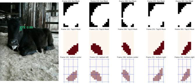
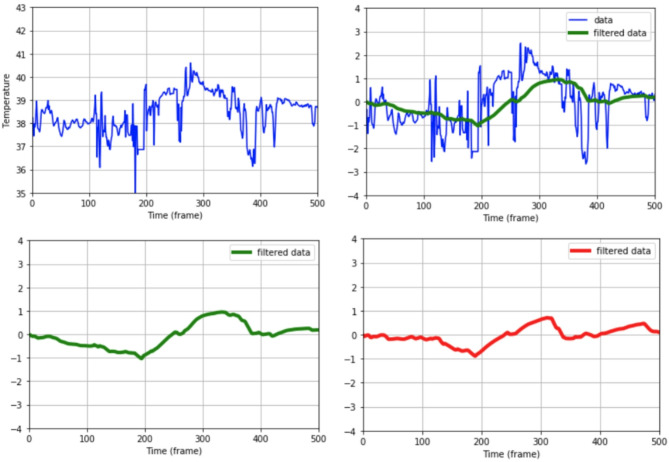
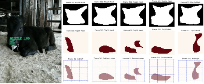
Methods: Thermal images were captured using a mobile infrared camera, and video recordings were obtained simultaneously from 11 calves. AI-based segmentation, utilizing previously trained weights, was used to automatically extract eye and muzzle ROIs from video images. 33 imaging sessions where the majority of frames exhibited reliable segmentation were selected for analysis. In Experiment 1, temperature data corresponding to the mean, top 10%, and top 30% values within each ROI underwent preprocessing steps (outlier rejection, standardization, and low-pass filtering) to derive temperature change patterns. This process generated six patterns per session (three for eyes and three muzzle regions), yielding a total of 198 patterns across all 33 image sessions. Cosine similarity analysis was then applied to quantify similarity within the same session. In Experiment 2, each ROI was divided into a 3 × 3 grid to map the distribution of high temperature values for spatial analysis. Statistical analyses included Kruskal-Wallis tests with Bonferroni corrections to assess regional differences.
Results: In Experiment 1, for the eyes, the patterns derived from the top 10% and 30% of temperatures had high cosine similarity (0.94). In contrast, the patterns based on the mean values had relatively lower similarities with the top 10% and 30% patterns (0.81 and 0.86, respectively). A similar trend was observed for the muzzle: the top 10% and 30% patterns had a high cosine similarity (0.93), while the patterns based on the mean values showed lower similarities (0.80, and 0.86). In Experiment 2, for the eyes, the top 10% of temperature values were mainly in the bottom region. In comparison, the top 30% of values were more evenly distributed in the mid and bottom regions. For the muzzles, the top 10% of temperature values were mainly distributed in both the top and bottom regions, and the top 30% of values were concentrated in the mid region.
Conclusion: This study demonstrates that integrating AI-based segmentation with infrared thermography enables precise identification of thermally reliable subregions within the eyes and muzzles of calves, leading to the extraction of temperature change patterns with high temporal consistency. The top 10% and 30% temperature values within these regions show higher pattern similarity than mean values, with distinct spatial distributions reflecting underlying vascular anatomy. Focusing on these high-temperature, vascularized subregions enhances the interpretability and reliability of temperature change pattern analysis for non-invasive monitoring of stress and physiological status in cattle, contributing to enhanced animal welfare.
期刊介绍:
BMC Veterinary Research is an open access, peer-reviewed journal that considers articles on all aspects of veterinary science and medicine, including the epidemiology, diagnosis, prevention and treatment of medical conditions of domestic, companion, farm and wild animals, as well as the biomedical processes that underlie their health.

 求助内容:
求助内容: 应助结果提醒方式:


