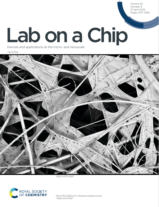
Fabrication of a bioreactor combining soft lithography and vat photopolymerisation to study tissues and multicellular organisms under dynamic culture conditions.
IF 6.1
2区 工程技术
Q1 BIOCHEMICAL RESEARCH METHODS
引用次数: 0
Abstract
Despite its capability to create much more realistic microenvironments for in vitro culturing of animal or human biological models, the spread of microfluidic tools in the world of biology and medicine has still not reached the predicted scale. Major obstacles to their widespread acceptance by end-users are manufacturing cost and operational complexity. 3D printing is a technology that is now widely democratised, thanks to its ease of use and very attractive cost/performance ratio. In particular, photopolymerisation through a liquid crystal screen is experiencing a very significant growth. Here, we describe the methodology we developed to evaluate this microfabrication technique and selected a photoprintable resin to manufacture a fluidic microsystem dedicated to tissue or micro-organism culture. The first originality of our approach lies in the architecture of the microsystem, which is made up of an elementary culture chamber in two parts, making it very easy to open after the culture period to carry out ex situ biochemical analyses. The second one is the nature of the materials used to make up the culture chamber, which consists of polydimethylsiloxane and a photoprinted resin. This hybrid assembly, combining an elastomer and a rigid plastic material, ensures a better seal and better dimensional control once the assembly is complete. We demonstrate the ability of our protocol to flexibly fabricate different culture chambers with dimensions down to 100 microns and we show for one of them three applications: a 2D layer of cell lines, a parasitic worm and a 3D microtissue from a pancreatic cancer patient.在动态培养条件下研究组织和多细胞生物的软光刻和还原光聚合相结合的生物反应器的制造。
尽管它能够为动物或人类生物模型的体外培养创造更现实的微环境,但微流体工具在生物学和医学领域的普及仍未达到预期的规模。最终用户广泛接受它们的主要障碍是制造成本和操作复杂性。3D打印是一项现在广泛普及的技术,这要归功于它的易用性和非常有吸引力的性价比。特别是,通过液晶屏的光聚合正经历着非常显著的增长。在这里,我们描述了我们开发的方法来评估这种微加工技术,并选择了一种光打印树脂来制造专门用于组织或微生物培养的流体微系统。我们的方法的第一个独创性在于微系统的结构,它由两个部分组成的初级培养室,使得它很容易在培养期后打开进行非原位生化分析。第二个是用来组成培养室的材料的性质,它由聚二甲基硅氧烷和光印树脂组成。这种混合组件结合了弹性体和刚性塑料材料,确保了更好的密封和更好的尺寸控制,一旦组装完成。我们展示了我们的方案能够灵活地制造尺寸低至100微米的不同培养室,我们展示了其中的三种应用之一:细胞系的二维层,寄生虫和来自胰腺癌患者的三维显微组织。
本文章由计算机程序翻译,如有差异,请以英文原文为准。
求助全文
约1分钟内获得全文
求助全文
来源期刊

Lab on a Chip
工程技术-化学综合
CiteScore
11.10
自引率
8.20%
发文量
434
审稿时长
2.6 months
期刊介绍:
Lab on a Chip is the premiere journal that publishes cutting-edge research in the field of miniaturization. By their very nature, microfluidic/nanofluidic/miniaturized systems are at the intersection of disciplines, spanning fundamental research to high-end application, which is reflected by the broad readership of the journal. Lab on a Chip publishes two types of papers on original research: full-length research papers and communications. Papers should demonstrate innovations, which can come from technical advancements or applications addressing pressing needs in globally important areas. The journal also publishes Comments, Reviews, and Perspectives.
 求助内容:
求助内容: 应助结果提醒方式:


