Janja Konjevod, Vanja Djukic, Tomislav Vukic, Klara Brekalo, Sara Abbaci Jangjel, Stefan Dimov, Rajko Fures, Vilmica Kapac, Mario Fuckar
{"title":"A Rare Case of Idiopathic Omental Necrosis in a Young Adult: Diagnostic Challenges and Management.","authors":"Janja Konjevod, Vanja Djukic, Tomislav Vukic, Klara Brekalo, Sara Abbaci Jangjel, Stefan Dimov, Rajko Fures, Vilmica Kapac, Mario Fuckar","doi":"10.5455/medarh.2025.79.237-240","DOIUrl":null,"url":null,"abstract":"<p><strong>Background: </strong>Idiopathic omental infarction (IOI) is a rare cause of acute abdominal pain that can clinically mimic more common conditions, such as acute appendicitis. IOI occurs due to vascular compromise of the greater omentum, leading to ischemia, pain, and often necrosis. Preoperative diagnosis remains challenging due to nonspecific clinical and laboratory findings, as well as the fact that clinicians rarely include IOI as a \"usual suspect\" in the differential diagnosis. Therefore, the condition is commonly identified intraoperatively.</p><p><strong>Objective: </strong>The aim of this article was to present a rare case of idipathic omental necrosis in young adult with description of appropriate diagnostic challanges and management.</p><p><strong>Methods: </strong>We present the case of a 23-year-old previously healthy male who was admitted for suspected acute appendicitis based on right lower quadrant pain, nausea, vomiting, and elevated inflammatory markers. Exploratory laparoscopy revealed no signs of appendicitis but identified hemoperitoneum and a necrotic segment of the greater omentum in its right upper segment. Appendectomy and resection of the infarcted omental tissue were performed. Histopathological analysis confirmed the diagnosis of omental infarction as well as the absence of histopathological signs of acute appendicitis.</p><p><strong>Conclusion: </strong>Idiopathic omental infarction, though rare, should be considered in the differential diagnosis of acute abdomen, particularly when clinical findings do not align with typical appendicitis. Advanced imaging modalities such as contrast-enhanced CT can facilitate preoperative diagnosis, potentially preventing unnecessary surgical interventions. However, in cases of diagnostic uncertainty, surgical exploration remains a mainstream approach. Awareness of this condition can improve diagnostic accuracy and optimize patient management.</p>","PeriodicalId":94135,"journal":{"name":"Medical archives (Sarajevo, Bosnia and Herzegovina)","volume":"79 3","pages":"237-240"},"PeriodicalIF":0.0000,"publicationDate":"2025-01-01","publicationTypes":"Journal Article","fieldsOfStudy":null,"isOpenAccess":false,"openAccessPdf":"https://www.ncbi.nlm.nih.gov/pmc/articles/PMC12253603/pdf/","citationCount":"0","resultStr":null,"platform":"Semanticscholar","paperid":null,"PeriodicalName":"Medical archives (Sarajevo, Bosnia and Herzegovina)","FirstCategoryId":"1085","ListUrlMain":"https://doi.org/10.5455/medarh.2025.79.237-240","RegionNum":0,"RegionCategory":null,"ArticlePicture":[],"TitleCN":null,"AbstractTextCN":null,"PMCID":null,"EPubDate":"","PubModel":"","JCR":"","JCRName":"","Score":null,"Total":0}
引用次数: 0
Abstract
Background: Idiopathic omental infarction (IOI) is a rare cause of acute abdominal pain that can clinically mimic more common conditions, such as acute appendicitis. IOI occurs due to vascular compromise of the greater omentum, leading to ischemia, pain, and often necrosis. Preoperative diagnosis remains challenging due to nonspecific clinical and laboratory findings, as well as the fact that clinicians rarely include IOI as a "usual suspect" in the differential diagnosis. Therefore, the condition is commonly identified intraoperatively.
Objective: The aim of this article was to present a rare case of idipathic omental necrosis in young adult with description of appropriate diagnostic challanges and management.
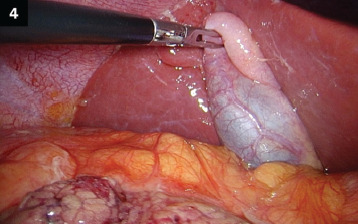
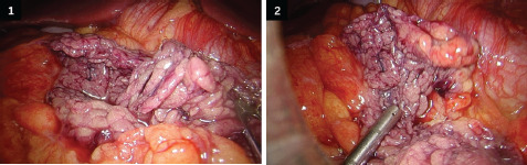
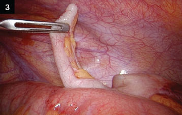
Methods: We present the case of a 23-year-old previously healthy male who was admitted for suspected acute appendicitis based on right lower quadrant pain, nausea, vomiting, and elevated inflammatory markers. Exploratory laparoscopy revealed no signs of appendicitis but identified hemoperitoneum and a necrotic segment of the greater omentum in its right upper segment. Appendectomy and resection of the infarcted omental tissue were performed. Histopathological analysis confirmed the diagnosis of omental infarction as well as the absence of histopathological signs of acute appendicitis.
Conclusion: Idiopathic omental infarction, though rare, should be considered in the differential diagnosis of acute abdomen, particularly when clinical findings do not align with typical appendicitis. Advanced imaging modalities such as contrast-enhanced CT can facilitate preoperative diagnosis, potentially preventing unnecessary surgical interventions. However, in cases of diagnostic uncertainty, surgical exploration remains a mainstream approach. Awareness of this condition can improve diagnostic accuracy and optimize patient management.

 求助内容:
求助内容: 应助结果提醒方式:


