Michal Březík, Veronika Matušková, Daniela Vysloužilová, Martin Šín, Veronika Chovancová, Kristína Sičová, Natália Jankaničová, Oldřich Chrapek
{"title":"Retinal Vessel Oxygen Saturation in Patients with Unilateral Edema of the Optic Nerve Head: A Pilot Study.","authors":"Michal Březík, Veronika Matušková, Daniela Vysloužilová, Martin Šín, Veronika Chovancová, Kristína Sičová, Natália Jankaničová, Oldřich Chrapek","doi":"10.2147/EB.S524274","DOIUrl":null,"url":null,"abstract":"<p><strong>Purpose: </strong>Evaluate whether optic disc edema results in changes in retinal microcirculation.</p><p><strong>Patients and methods: </strong>The study group consisted of 11 patients with unilateral optic disc edema (papilledema). The control group consisted of the healthy eyes of the same 11 patients. Patients underwent non-invasive photo-spectrometric retinal oximetry using Oxymap T1 retinal oximeter (Oxymap, Reykjavik, Iceland). In the eyes of these 11 patients, we examined the diameter of the retinal arteries and veins, arterial and venous blood oxygen saturation, and the difference in oxygen saturation between arterioles and venules (A-V difference).</p><p><strong>Results: </strong>In the papilledema group, the decrease in the retinal arterial diameter was statistically significant (p=0.001). The median diameter of the retinal artery was 11.70 px (IQR 1.47) or after conversion 109.00 µm (IQR 14.00) in the papilledema group and 13.75 px (IQR 1.61) or 128.00 µm (IQR 15.00) in the control group. The increase in the diameter of the retinal veins in the papilledema group was statistically significant (p=0.012), where the median diameter in the papilledema group was 20.88 px (IQR 3.72) or 194.00 µm (IQR 35.00), and in the control group was 18.18 px (IQR 3.60) or 169 µm (IQR 33.00). There was a statistically significant decrease (p<0.001) in the venous saturation in the papilledema group with a median value of 53.16% (IQR 17.38) and 60.02% (IQR 11.98) in the control group. The median of the A-V difference was 51.92 (IQR 15.96) in the papilledema group, resp. 38.49 (IQR 9.75) in the control group and a significant increase in the papilledema group (p<0.001) was reported.</p><p><strong>Conclusion: </strong>Automatic retinal oximetry demonstrated changes in the retinal microcirculation in patients with optic disc edema.</p>","PeriodicalId":51844,"journal":{"name":"Eye and Brain","volume":"17 ","pages":"81-94"},"PeriodicalIF":2.4000,"publicationDate":"2025-07-08","publicationTypes":"Journal Article","fieldsOfStudy":null,"isOpenAccess":false,"openAccessPdf":"https://www.ncbi.nlm.nih.gov/pmc/articles/PMC12255252/pdf/","citationCount":"0","resultStr":null,"platform":"Semanticscholar","paperid":null,"PeriodicalName":"Eye and Brain","FirstCategoryId":"1085","ListUrlMain":"https://doi.org/10.2147/EB.S524274","RegionNum":0,"RegionCategory":null,"ArticlePicture":[],"TitleCN":null,"AbstractTextCN":null,"PMCID":null,"EPubDate":"2025/1/1 0:00:00","PubModel":"eCollection","JCR":"Q1","JCRName":"OPHTHALMOLOGY","Score":null,"Total":0}
引用次数: 0
Abstract
Purpose: Evaluate whether optic disc edema results in changes in retinal microcirculation.
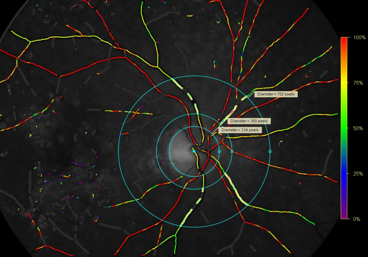
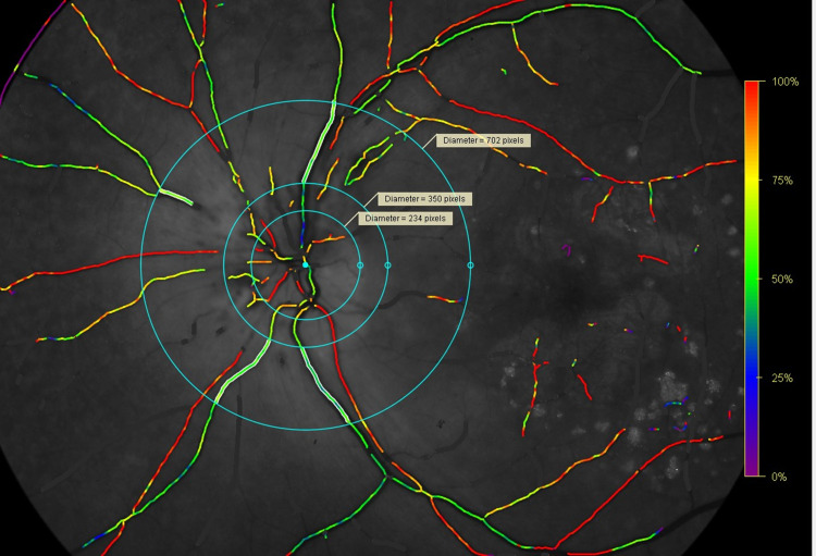
Patients and methods: The study group consisted of 11 patients with unilateral optic disc edema (papilledema). The control group consisted of the healthy eyes of the same 11 patients. Patients underwent non-invasive photo-spectrometric retinal oximetry using Oxymap T1 retinal oximeter (Oxymap, Reykjavik, Iceland). In the eyes of these 11 patients, we examined the diameter of the retinal arteries and veins, arterial and venous blood oxygen saturation, and the difference in oxygen saturation between arterioles and venules (A-V difference).
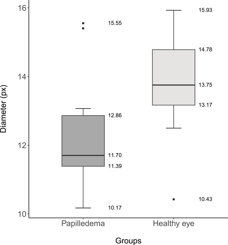
Results: In the papilledema group, the decrease in the retinal arterial diameter was statistically significant (p=0.001). The median diameter of the retinal artery was 11.70 px (IQR 1.47) or after conversion 109.00 µm (IQR 14.00) in the papilledema group and 13.75 px (IQR 1.61) or 128.00 µm (IQR 15.00) in the control group. The increase in the diameter of the retinal veins in the papilledema group was statistically significant (p=0.012), where the median diameter in the papilledema group was 20.88 px (IQR 3.72) or 194.00 µm (IQR 35.00), and in the control group was 18.18 px (IQR 3.60) or 169 µm (IQR 33.00). There was a statistically significant decrease (p<0.001) in the venous saturation in the papilledema group with a median value of 53.16% (IQR 17.38) and 60.02% (IQR 11.98) in the control group. The median of the A-V difference was 51.92 (IQR 15.96) in the papilledema group, resp. 38.49 (IQR 9.75) in the control group and a significant increase in the papilledema group (p<0.001) was reported.
Conclusion: Automatic retinal oximetry demonstrated changes in the retinal microcirculation in patients with optic disc edema.
期刊介绍:
Eye and Brain is an international, peer-reviewed, open access journal focusing on basic research, clinical findings, and expert reviews in the field of visual science and neuro-ophthalmology. The journal’s unique focus is the link between two well-known visual centres, the eye and the brain, with an emphasis on the importance of such connections. All aspects of clinical and especially basic research on the visual system are addressed within the journal as well as significant future directions in vision research and therapeutic measures. This unique journal focuses on neurological aspects of vision – both physiological and pathological. The scope of the journal spans from the cornea to the associational visual cortex and all the visual centers in between. Topics range from basic biological mechanisms to therapeutic treatment, from simple organisms to humans, and utilizing techniques from molecular biology to behavior. The journal especially welcomes primary research articles or review papers that make the connection between the eye and the brain. Specific areas covered in the journal include: Physiology and pathophysiology of visual centers, Eye movement disorders and strabismus, Cellular, biochemical, and molecular features of the visual system, Structural and functional organization of the eye and of the visual cortex, Metabolic demands of the visual system, Diseases and disorders with neuro-ophthalmic manifestations, Clinical and experimental neuro-ophthalmology and visual system pathologies, Epidemiological studies.

 求助内容:
求助内容: 应助结果提醒方式:


