Teja Pathour, Ghazal Rastegar, Shashank R Sirsi, Baowei Fei
{"title":"Harnessing chemically crosslinked microbubble clusters using deep learning for ultrasound contrast imaging.","authors":"Teja Pathour, Ghazal Rastegar, Shashank R Sirsi, Baowei Fei","doi":"10.1117/1.JMI.12.4.047001","DOIUrl":null,"url":null,"abstract":"<p><strong>Purpose: </strong>We aim to investigate and isolate the distinctive acoustic properties generated by chemically crosslinked microbubble clusters (CCMCs) using machine learning (ML) techniques, specifically using an anomaly detection model based on autoencoders.</p><p><strong>Approach: </strong>CCMCs were synthesized via copper-free click chemistry and subjected to acoustic analysis using a clinical transducer. Radiofrequency data were acquired, processed, and organized into training and testing datasets for the ML models. We trained an anomaly detection model with the nonclustered microbubbles (MBs) and tested the model on the CCMCs to isolate the unique acoustics. We also had a separate set of control experiments that was performed to validate the anomaly detection model.</p><p><strong>Results: </strong>The anomaly detection model successfully identified frames exhibiting unique acoustic signatures associated with CCMCs. Frequency domain analysis further confirmed that these frames displayed higher amplitude and energy, suggesting the occurrence of potential coalescence events. The specificity of the model was validated through control experiments, in which both groups contained only individual MBs without clustering. As anticipated, no anomalies were detected in this control dataset, reinforcing the model's ability to distinguish clustered MBs from nonclustered ones.</p><p><strong>Conclusions: </strong>We highlight the feasibility of detecting and distinguishing the unique acoustic characteristics of CCMCs, thereby improving the detectability and localization of contrast agents in ultrasound imaging. The elevated acoustic amplitudes produced by CCMCs offer potential advantages for more effective contrast agent detection, which is particularly valuable in super-resolution ultrasound imaging. Both the contrast agent and the ML-based analysis approach hold promise for a wide range of applications.</p>","PeriodicalId":47707,"journal":{"name":"Journal of Medical Imaging","volume":"12 4","pages":"047001"},"PeriodicalIF":1.7000,"publicationDate":"2025-07-01","publicationTypes":"Journal Article","fieldsOfStudy":null,"isOpenAccess":false,"openAccessPdf":"https://www.ncbi.nlm.nih.gov/pmc/articles/PMC12255354/pdf/","citationCount":"0","resultStr":null,"platform":"Semanticscholar","paperid":null,"PeriodicalName":"Journal of Medical Imaging","FirstCategoryId":"3","ListUrlMain":"https://doi.org/10.1117/1.JMI.12.4.047001","RegionNum":0,"RegionCategory":null,"ArticlePicture":[],"TitleCN":null,"AbstractTextCN":null,"PMCID":null,"EPubDate":"2025/7/12 0:00:00","PubModel":"Epub","JCR":"Q3","JCRName":"RADIOLOGY, NUCLEAR MEDICINE & MEDICAL IMAGING","Score":null,"Total":0}
引用次数: 0
Abstract
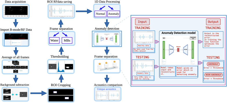
Purpose: We aim to investigate and isolate the distinctive acoustic properties generated by chemically crosslinked microbubble clusters (CCMCs) using machine learning (ML) techniques, specifically using an anomaly detection model based on autoencoders.
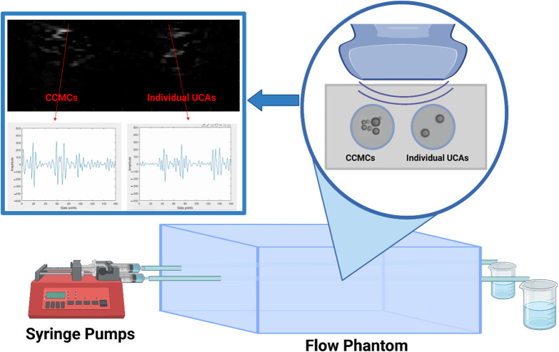
Approach: CCMCs were synthesized via copper-free click chemistry and subjected to acoustic analysis using a clinical transducer. Radiofrequency data were acquired, processed, and organized into training and testing datasets for the ML models. We trained an anomaly detection model with the nonclustered microbubbles (MBs) and tested the model on the CCMCs to isolate the unique acoustics. We also had a separate set of control experiments that was performed to validate the anomaly detection model.
Results: The anomaly detection model successfully identified frames exhibiting unique acoustic signatures associated with CCMCs. Frequency domain analysis further confirmed that these frames displayed higher amplitude and energy, suggesting the occurrence of potential coalescence events. The specificity of the model was validated through control experiments, in which both groups contained only individual MBs without clustering. As anticipated, no anomalies were detected in this control dataset, reinforcing the model's ability to distinguish clustered MBs from nonclustered ones.
Conclusions: We highlight the feasibility of detecting and distinguishing the unique acoustic characteristics of CCMCs, thereby improving the detectability and localization of contrast agents in ultrasound imaging. The elevated acoustic amplitudes produced by CCMCs offer potential advantages for more effective contrast agent detection, which is particularly valuable in super-resolution ultrasound imaging. Both the contrast agent and the ML-based analysis approach hold promise for a wide range of applications.
期刊介绍:
JMI covers fundamental and translational research, as well as applications, focused on medical imaging, which continue to yield physical and biomedical advancements in the early detection, diagnostics, and therapy of disease as well as in the understanding of normal. The scope of JMI includes: Imaging physics, Tomographic reconstruction algorithms (such as those in CT and MRI), Image processing and deep learning, Computer-aided diagnosis and quantitative image analysis, Visualization and modeling, Picture archiving and communications systems (PACS), Image perception and observer performance, Technology assessment, Ultrasonic imaging, Image-guided procedures, Digital pathology, Biomedical applications of biomedical imaging. JMI allows for the peer-reviewed communication and archiving of scientific developments, translational and clinical applications, reviews, and recommendations for the field.

 求助内容:
求助内容: 应助结果提醒方式:


