Diagnostic cut-off values and grading of carpal tunnel syndrome by shear wave elastography at different tunnel locations correlated with gold standard nerve conduction study - a case-control study.
IF 1.5 Q3 RADIOLOGY, NUCLEAR MEDICINE & MEDICAL IMAGING
{"title":"Diagnostic cut-off values and grading of carpal tunnel syndrome by shear wave elastography at different tunnel locations correlated with gold standard nerve conduction study - a case-control study.","authors":"Prashat Bhalke, Priya Pattath Sankaran, Arvind N Prabhu, Rajagopal Kadavigere, Prakashini Koteshwara","doi":"10.15557/jou.2025.0017","DOIUrl":null,"url":null,"abstract":"<p><strong>Aim: </strong>The gold standard nerve conduction study for diagnosing carpal tunnel syndrome is often painful and has variable diagnostic accuracy. This study aimed to evaluate the diagnostic performance of shear wave elastography in correlation with nerve conduction study.</p><p><strong>Material and methods: </strong>A prospective case-control study was conducted on 50 participants (50 wrists), including 25 carpal tunnel syndrome cases diagnosed by nerve conduction study and 25 healthy controls. Shear wave elastography assessed the stiffness of the median nerve at three locations: outside the carpal tunnel, at the inlet, and at the outlet. Cross-sectional area measurements were also obtained using B-mode ultrasound. Receiver operating characteristic curves were used to evaluate diagnostic performance.</p><p><strong>Results: </strong>Shear wave elastography and cross-sectional area demonstrated high diagnostic accuracy for carpal tunnel syndrome, with a cut-off value of ≥63.5 kPa inside the tunnel (mean of inlet and outlet values) and a cross-sectional area cut-off of ≥0.08 cm<sup>2</sup> at the inlet of the tunnel offering optimal performance. While cross-sectional area provided high sensitivity, shear wave elastography showed superior specificity; their combination improved overall diagnostic accuracy. Shear wave elastography values did not significantly differ across carpal tunnel syndrome severity grades based on nerve conduction study (<i>p</i> >0.05). However, shear wave elastography at the tunnel inlet differentiated severe carpal tunnel syndrome from non-severe cases (<i>p</i> = 0.045), with a cut-off of ≥126 kPa predicting severe carpal tunnel syndrome with 100% sensitivity, 77% specificity, and an area under the receiver operating characteristic curve of 0.871.</p><p><strong>Conclusions: </strong>Shear wave elastography is a reliable, non-invasive modality for carpal tunnel syndrome diagnosis, offering excellent specificity, particularly when combined with cross-sectional area. Additionally, shear wave elastography at the tunnel inlet may help identify severe carpal tunnel syndrome, supporting timely clinical decision-making and prioritization of intervention.</p>","PeriodicalId":45612,"journal":{"name":"Journal of Ultrasonography","volume":"25 101","pages":"20250017"},"PeriodicalIF":1.5000,"publicationDate":"2025-06-30","publicationTypes":"Journal Article","fieldsOfStudy":null,"isOpenAccess":false,"openAccessPdf":"https://www.ncbi.nlm.nih.gov/pmc/articles/PMC12255475/pdf/","citationCount":"0","resultStr":null,"platform":"Semanticscholar","paperid":null,"PeriodicalName":"Journal of Ultrasonography","FirstCategoryId":"1085","ListUrlMain":"https://doi.org/10.15557/jou.2025.0017","RegionNum":0,"RegionCategory":null,"ArticlePicture":[],"TitleCN":null,"AbstractTextCN":null,"PMCID":null,"EPubDate":"2025/4/1 0:00:00","PubModel":"eCollection","JCR":"Q3","JCRName":"RADIOLOGY, NUCLEAR MEDICINE & MEDICAL IMAGING","Score":null,"Total":0}
引用次数: 0
Abstract
Aim: The gold standard nerve conduction study for diagnosing carpal tunnel syndrome is often painful and has variable diagnostic accuracy. This study aimed to evaluate the diagnostic performance of shear wave elastography in correlation with nerve conduction study.
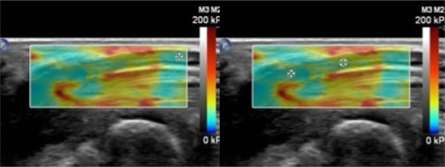
Material and methods: A prospective case-control study was conducted on 50 participants (50 wrists), including 25 carpal tunnel syndrome cases diagnosed by nerve conduction study and 25 healthy controls. Shear wave elastography assessed the stiffness of the median nerve at three locations: outside the carpal tunnel, at the inlet, and at the outlet. Cross-sectional area measurements were also obtained using B-mode ultrasound. Receiver operating characteristic curves were used to evaluate diagnostic performance.
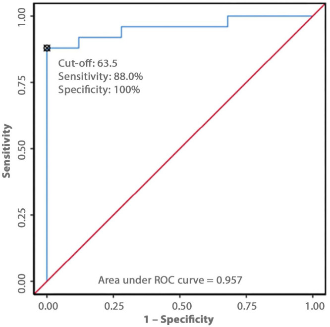
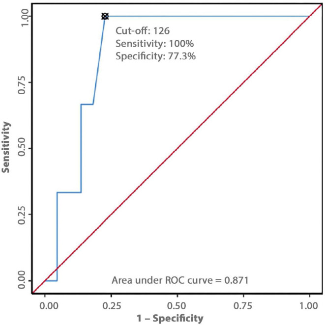
Results: Shear wave elastography and cross-sectional area demonstrated high diagnostic accuracy for carpal tunnel syndrome, with a cut-off value of ≥63.5 kPa inside the tunnel (mean of inlet and outlet values) and a cross-sectional area cut-off of ≥0.08 cm2 at the inlet of the tunnel offering optimal performance. While cross-sectional area provided high sensitivity, shear wave elastography showed superior specificity; their combination improved overall diagnostic accuracy. Shear wave elastography values did not significantly differ across carpal tunnel syndrome severity grades based on nerve conduction study (p >0.05). However, shear wave elastography at the tunnel inlet differentiated severe carpal tunnel syndrome from non-severe cases (p = 0.045), with a cut-off of ≥126 kPa predicting severe carpal tunnel syndrome with 100% sensitivity, 77% specificity, and an area under the receiver operating characteristic curve of 0.871.
Conclusions: Shear wave elastography is a reliable, non-invasive modality for carpal tunnel syndrome diagnosis, offering excellent specificity, particularly when combined with cross-sectional area. Additionally, shear wave elastography at the tunnel inlet may help identify severe carpal tunnel syndrome, supporting timely clinical decision-making and prioritization of intervention.

 求助内容:
求助内容: 应助结果提醒方式:


