Sławomir Witkowski, Maria Respondek-Liberska, Iwona Strzelecka
{"title":"Hypoplastic left heart syndrome with mitral regurgitation: a new marker of poor fetal outcome.","authors":"Sławomir Witkowski, Maria Respondek-Liberska, Iwona Strzelecka","doi":"10.15557/jou.2025.0009","DOIUrl":null,"url":null,"abstract":"<p><strong>Aim: </strong>Hypoplastic left heart syndrome is a severe congenital heart defect that may be accompanied by tricuspid and mitral valve regurgitation. The aim of this paper is to identify a new ultrasound marker for evaluating the outcomes of fetuses with hypoplastic left heart syndrome.</p><p><strong>Material and methods: </strong>This was a single-center analysis of a group of fetuses who underwent fetal ultrasound examinations at our tertiary center between 2016 and 2023. This study included 80 fetuses with hypoplastic left heart syndrome. The gestational age of the studied fetuses ranged from 16.3 to 39.5 weeks. All anomalies and irregularities accompanying hypoplastic left heart syndrome were detected during the second and third trimesters of pregnancy.</p><p><strong>Results: </strong>Among fetuses with hypoplastic left heart syndrome with tricuspid regurgitation, the mortality rate was 0% (0/16) and the survival rate was 100% (16/16). In contrast, in fetuses with hypoplastic left heart syndrome with both tricuspid and mitral regurgitation, the mortality rate was 75% (3/4) and the survival rate was 25% (1/4). The incidence of death was significantly higher in the group of fetuses with hypoplastic left heart syndrome with both tricuspid and mitral regurgitation compared to the group with tricuspid regurgitation (Yates's chi-squared test: <i>p</i> = 0.003; Fisher's test: <i>p</i> = 0.0035).</p><p><strong>Conclusions: </strong>The coexistence of hypoplastic left heart syndrome with tricuspid and mitral regurgitation is significantly associated with the death of newborns even when treatment and/or surgery is performed. Therefore, the presence of mitral regurgitation in fetuses with hypoplastic left heart syndrome may serve as an additional ultrasound marker for poor neonatal outcome.</p>","PeriodicalId":45612,"journal":{"name":"Journal of Ultrasonography","volume":"25 100","pages":"20250009"},"PeriodicalIF":1.5000,"publicationDate":"2025-04-23","publicationTypes":"Journal Article","fieldsOfStudy":null,"isOpenAccess":false,"openAccessPdf":"https://www.ncbi.nlm.nih.gov/pmc/articles/PMC12246897/pdf/","citationCount":"0","resultStr":null,"platform":"Semanticscholar","paperid":null,"PeriodicalName":"Journal of Ultrasonography","FirstCategoryId":"1085","ListUrlMain":"https://doi.org/10.15557/jou.2025.0009","RegionNum":0,"RegionCategory":null,"ArticlePicture":[],"TitleCN":null,"AbstractTextCN":null,"PMCID":null,"EPubDate":"2025/1/1 0:00:00","PubModel":"eCollection","JCR":"Q3","JCRName":"RADIOLOGY, NUCLEAR MEDICINE & MEDICAL IMAGING","Score":null,"Total":0}
引用次数: 0
Abstract
Aim: Hypoplastic left heart syndrome is a severe congenital heart defect that may be accompanied by tricuspid and mitral valve regurgitation. The aim of this paper is to identify a new ultrasound marker for evaluating the outcomes of fetuses with hypoplastic left heart syndrome.
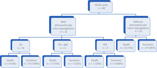
Material and methods: This was a single-center analysis of a group of fetuses who underwent fetal ultrasound examinations at our tertiary center between 2016 and 2023. This study included 80 fetuses with hypoplastic left heart syndrome. The gestational age of the studied fetuses ranged from 16.3 to 39.5 weeks. All anomalies and irregularities accompanying hypoplastic left heart syndrome were detected during the second and third trimesters of pregnancy.
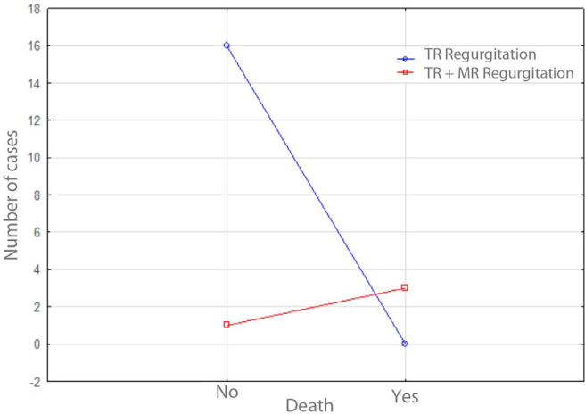
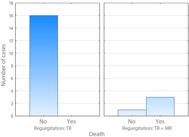
Results: Among fetuses with hypoplastic left heart syndrome with tricuspid regurgitation, the mortality rate was 0% (0/16) and the survival rate was 100% (16/16). In contrast, in fetuses with hypoplastic left heart syndrome with both tricuspid and mitral regurgitation, the mortality rate was 75% (3/4) and the survival rate was 25% (1/4). The incidence of death was significantly higher in the group of fetuses with hypoplastic left heart syndrome with both tricuspid and mitral regurgitation compared to the group with tricuspid regurgitation (Yates's chi-squared test: p = 0.003; Fisher's test: p = 0.0035).
Conclusions: The coexistence of hypoplastic left heart syndrome with tricuspid and mitral regurgitation is significantly associated with the death of newborns even when treatment and/or surgery is performed. Therefore, the presence of mitral regurgitation in fetuses with hypoplastic left heart syndrome may serve as an additional ultrasound marker for poor neonatal outcome.

 求助内容:
求助内容: 应助结果提醒方式:


