Hamzeh Yacoub, Aya Aqel, Mohammed Adas, Qais Hjouj, Zaid Yacoub, Rita Yacoub, Hadi Dababseh
{"title":"Extremely Rare Presentation of Pilonidal Sinus Disease in the Posterior Cranial Fossa of a 2-Year-Old Patient: A Case Report.","authors":"Hamzeh Yacoub, Aya Aqel, Mohammed Adas, Qais Hjouj, Zaid Yacoub, Rita Yacoub, Hadi Dababseh","doi":"10.1055/a-2641-6301","DOIUrl":null,"url":null,"abstract":"<p><p>A 2-year-old female patient presented after experiencing a generalized tonic-clonic seizure accompanied by fever, followed by a loss of consciousness. She underwent an urgent right frontal external ventricular drain placement. Intraoperative cerebrospinal fluid analysis was negative for infectious patterns. MRI showed a predominantly cystic lesion in the midline posterior fossa, with a compressive mass effect. Subsequently, she underwent a suboccipital craniotomy for microscopic resection of a posterior cranial fossa lesion. Histopathology reported keratin flakes with severe active inflammation, and foreign body type giant cell reaction in scalp excision with free hair shafts through the inflammatory focus, consistent with pilonidal sinus. The patient was then discharged home in good health.</p>","PeriodicalId":44256,"journal":{"name":"Journal of Neurological Surgery Reports","volume":"86 3","pages":"e149-e152"},"PeriodicalIF":0.7000,"publicationDate":"2025-07-11","publicationTypes":"Journal Article","fieldsOfStudy":null,"isOpenAccess":false,"openAccessPdf":"https://www.ncbi.nlm.nih.gov/pmc/articles/PMC12254008/pdf/","citationCount":"0","resultStr":null,"platform":"Semanticscholar","paperid":null,"PeriodicalName":"Journal of Neurological Surgery Reports","FirstCategoryId":"1085","ListUrlMain":"https://doi.org/10.1055/a-2641-6301","RegionNum":0,"RegionCategory":null,"ArticlePicture":[],"TitleCN":null,"AbstractTextCN":null,"PMCID":null,"EPubDate":"2025/7/1 0:00:00","PubModel":"eCollection","JCR":"Q4","JCRName":"CLINICAL NEUROLOGY","Score":null,"Total":0}
引用次数: 0
Abstract
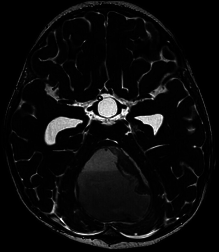
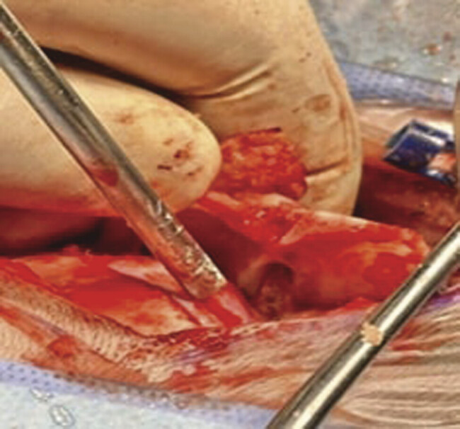
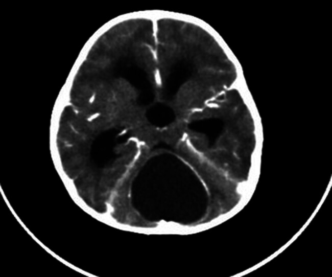
A 2-year-old female patient presented after experiencing a generalized tonic-clonic seizure accompanied by fever, followed by a loss of consciousness. She underwent an urgent right frontal external ventricular drain placement. Intraoperative cerebrospinal fluid analysis was negative for infectious patterns. MRI showed a predominantly cystic lesion in the midline posterior fossa, with a compressive mass effect. Subsequently, she underwent a suboccipital craniotomy for microscopic resection of a posterior cranial fossa lesion. Histopathology reported keratin flakes with severe active inflammation, and foreign body type giant cell reaction in scalp excision with free hair shafts through the inflammatory focus, consistent with pilonidal sinus. The patient was then discharged home in good health.

 求助内容:
求助内容: 应助结果提醒方式:


