Lubna S Khan, Sifa Farheen, Vijay K Pandey, Vinay P Singh, Manoj G Madakshira
{"title":"Chondromyxoid Fibroma of Phalanges.","authors":"Lubna S Khan, Sifa Farheen, Vijay K Pandey, Vinay P Singh, Manoj G Madakshira","doi":"10.18295/squmj.10.2024.068","DOIUrl":null,"url":null,"abstract":"<p><p>Chondromyxoid fibroma (CMF) is a rare, benign metaphyseal bone tumour characterised by a combination of chondroid, myxoid, and fibrous elements. It can affect any bone at any age, with no gender predilection. We report a case of CMF in a 47-year-old female patient who presented with swelling of the left little finger at a tertiary care hospital in Kolkata, India, in 2024. Imaging revealed a lobulated lesion originating from the middle phalanx of the left little finger, extending to involve the base of the distal phalanx. Fine needle aspiration cytology indicated a moderately cellular myxoid lesion. Histopathology, which remains crucial for diagnosis, showed a lobulated tumour with zones of spindle to stellate cells, associated with an abundant myxoid and chondroid matrix in the intercellular spaces. Treatment involves complete local excision with tumour-free margins, as recurrence may occur with local curettage.</p>","PeriodicalId":22083,"journal":{"name":"Sultan Qaboos University Medical Journal","volume":"25 1","pages":"168-174"},"PeriodicalIF":0.0000,"publicationDate":"2025-05-02","publicationTypes":"Journal Article","fieldsOfStudy":null,"isOpenAccess":false,"openAccessPdf":"https://www.ncbi.nlm.nih.gov/pmc/articles/PMC12255334/pdf/","citationCount":"0","resultStr":null,"platform":"Semanticscholar","paperid":null,"PeriodicalName":"Sultan Qaboos University Medical Journal","FirstCategoryId":"1085","ListUrlMain":"https://doi.org/10.18295/squmj.10.2024.068","RegionNum":0,"RegionCategory":null,"ArticlePicture":[],"TitleCN":null,"AbstractTextCN":null,"PMCID":null,"EPubDate":"2025/1/1 0:00:00","PubModel":"eCollection","JCR":"Q3","JCRName":"Medicine","Score":null,"Total":0}
引用次数: 0
Abstract
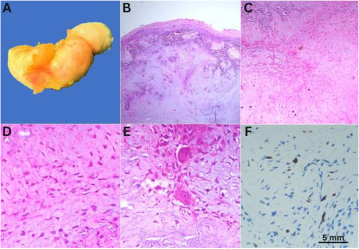
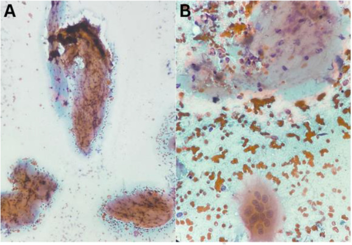
Chondromyxoid fibroma (CMF) is a rare, benign metaphyseal bone tumour characterised by a combination of chondroid, myxoid, and fibrous elements. It can affect any bone at any age, with no gender predilection. We report a case of CMF in a 47-year-old female patient who presented with swelling of the left little finger at a tertiary care hospital in Kolkata, India, in 2024. Imaging revealed a lobulated lesion originating from the middle phalanx of the left little finger, extending to involve the base of the distal phalanx. Fine needle aspiration cytology indicated a moderately cellular myxoid lesion. Histopathology, which remains crucial for diagnosis, showed a lobulated tumour with zones of spindle to stellate cells, associated with an abundant myxoid and chondroid matrix in the intercellular spaces. Treatment involves complete local excision with tumour-free margins, as recurrence may occur with local curettage.


 求助内容:
求助内容: 应助结果提醒方式:


