Utilizing of Seaweed for Eco-friendly Synthesis of Nickel Cobalt Nanoparticles and Assessment of its Cytotoxic Effects on Human Liver and Colon Cancer Cells.
Eman A Alanazy, Daoud Ali, Mohammed H A Almarzoug, Khadijah N Yaseen, Bader O Almutairi, Saad Alkahtani, Badr A Aldahmash, Saud Alarifi
{"title":"Utilizing of Seaweed for Eco-friendly Synthesis of Nickel Cobalt Nanoparticles and Assessment of its Cytotoxic Effects on Human Liver and Colon Cancer Cells.","authors":"Eman A Alanazy, Daoud Ali, Mohammed H A Almarzoug, Khadijah N Yaseen, Bader O Almutairi, Saad Alkahtani, Badr A Aldahmash, Saud Alarifi","doi":"10.1177/15593258251360056","DOIUrl":null,"url":null,"abstract":"<p><p>In this study we used biosynthesis methods to create bimetallic nickel cobalt nanoparticles (Ni-Co NPs) utilizing seaweed. Before exposure to target cells, the characterization of Ni-Co NPs is done by UV-Vis spectrophotometry, EDX, SEM, TEM, the shape of g Ni-Co NPs are polygonal form and its size is measured 38.27 ± 3 nm. The cytotoxic effect of g Ni-Co NPs on HuH7 and HCT cells were determined by MTT and NRU assays. The cytotoxicity of NPs increased in a concentration dependent manner and it showed high cytotoxic effect on HCT-116 cells than HuH-7 cells. We determined IC<sub>50</sub> 24 h for HuH-7 and HCT -116 cells at 24 h, it was 65.84 and 24.73 μg/mL, respectively. ROS was elevated at higher concentration of Ni-Co NPs. LPO was increased at 16 μg/mL in HuH-7 cells and 19 μg/mL in HCT-116 cells. CAT was reduced in HCT-116 cells than HuH-7 cells high concentration of NPs. JC-1 staining, the loss of MMP in control, Ni-Co NPs exposed cell were evaluated. In HuH-7 and HCT-116 cells, maximum apoptotic cells were observed at high concentration. Apoptotic genes were expressed in both type cells. The above findings highlight the significance of Ni-Co NPs and useful in a number of cancer treatments.</p>","PeriodicalId":11285,"journal":{"name":"Dose-Response","volume":"23 3","pages":"15593258251360056"},"PeriodicalIF":2.4000,"publicationDate":"2025-07-11","publicationTypes":"Journal Article","fieldsOfStudy":null,"isOpenAccess":false,"openAccessPdf":"https://www.ncbi.nlm.nih.gov/pmc/articles/PMC12254618/pdf/","citationCount":"0","resultStr":null,"platform":"Semanticscholar","paperid":null,"PeriodicalName":"Dose-Response","FirstCategoryId":"3","ListUrlMain":"https://doi.org/10.1177/15593258251360056","RegionNum":4,"RegionCategory":"医学","ArticlePicture":[],"TitleCN":null,"AbstractTextCN":null,"PMCID":null,"EPubDate":"2025/7/1 0:00:00","PubModel":"eCollection","JCR":"Q3","JCRName":"PHARMACOLOGY & PHARMACY","Score":null,"Total":0}
引用次数: 0
Abstract
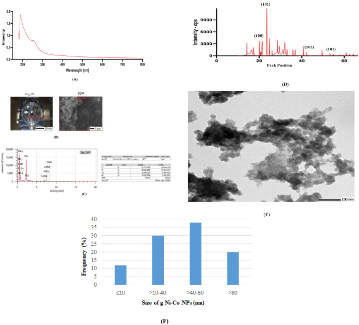
In this study we used biosynthesis methods to create bimetallic nickel cobalt nanoparticles (Ni-Co NPs) utilizing seaweed. Before exposure to target cells, the characterization of Ni-Co NPs is done by UV-Vis spectrophotometry, EDX, SEM, TEM, the shape of g Ni-Co NPs are polygonal form and its size is measured 38.27 ± 3 nm. The cytotoxic effect of g Ni-Co NPs on HuH7 and HCT cells were determined by MTT and NRU assays. The cytotoxicity of NPs increased in a concentration dependent manner and it showed high cytotoxic effect on HCT-116 cells than HuH-7 cells. We determined IC50 24 h for HuH-7 and HCT -116 cells at 24 h, it was 65.84 and 24.73 μg/mL, respectively. ROS was elevated at higher concentration of Ni-Co NPs. LPO was increased at 16 μg/mL in HuH-7 cells and 19 μg/mL in HCT-116 cells. CAT was reduced in HCT-116 cells than HuH-7 cells high concentration of NPs. JC-1 staining, the loss of MMP in control, Ni-Co NPs exposed cell were evaluated. In HuH-7 and HCT-116 cells, maximum apoptotic cells were observed at high concentration. Apoptotic genes were expressed in both type cells. The above findings highlight the significance of Ni-Co NPs and useful in a number of cancer treatments.
Dose-ResponsePHARMACOLOGY & PHARMACY-RADIOLOGY, NUCLEAR MEDICINE & MEDICAL IMAGING
CiteScore
4.90
自引率
4.00%
发文量
140
审稿时长
>12 weeks
期刊介绍:
Dose-Response is an open access peer-reviewed online journal publishing original findings and commentaries on the occurrence of dose-response relationships across a broad range of disciplines. Particular interest focuses on experimental evidence providing mechanistic understanding of nonlinear dose-response relationships.


 求助内容:
求助内容: 应助结果提醒方式:


