Dejan M Rašić, Dolika D Vasović, Miroslav Knežević
{"title":"Primary Orbital Teratoma With Congenital Anophthalmia in a Neonate: A Rare Case With Histopathological and Radiological Correlation.","authors":"Dejan M Rašić, Dolika D Vasović, Miroslav Knežević","doi":"10.1155/crop/5032089","DOIUrl":null,"url":null,"abstract":"<p><p>This case report describes a rare instance of primary orbital teratoma with anophthalmia in a neonate. A 6-day-old female presented with a congenital right orbital swelling and absence of visible ocular structures. MRI revealed a large, well-vascularized orbital mass without intracranial extension, accompanied by malformations in the right cerebral hemisphere. Histopathological examination confirmed a benign, mature/mixed teratoma comprising elements from all three germ layers, including neuroectoderm, mesoderm, and endoderm, with no evidence of malignancy. The patient underwent successful orbital exenteration with an implant at 3 weeks of age.</p>","PeriodicalId":9603,"journal":{"name":"Case Reports in Ophthalmological Medicine","volume":"2025 ","pages":"5032089"},"PeriodicalIF":0.4000,"publicationDate":"2025-06-27","publicationTypes":"Journal Article","fieldsOfStudy":null,"isOpenAccess":false,"openAccessPdf":"https://www.ncbi.nlm.nih.gov/pmc/articles/PMC12253985/pdf/","citationCount":"0","resultStr":null,"platform":"Semanticscholar","paperid":null,"PeriodicalName":"Case Reports in Ophthalmological Medicine","FirstCategoryId":"1085","ListUrlMain":"https://doi.org/10.1155/crop/5032089","RegionNum":0,"RegionCategory":null,"ArticlePicture":[],"TitleCN":null,"AbstractTextCN":null,"PMCID":null,"EPubDate":"2025/1/1 0:00:00","PubModel":"eCollection","JCR":"Q4","JCRName":"OPHTHALMOLOGY","Score":null,"Total":0}
引用次数: 0
Abstract
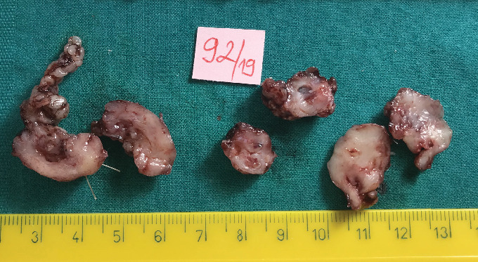
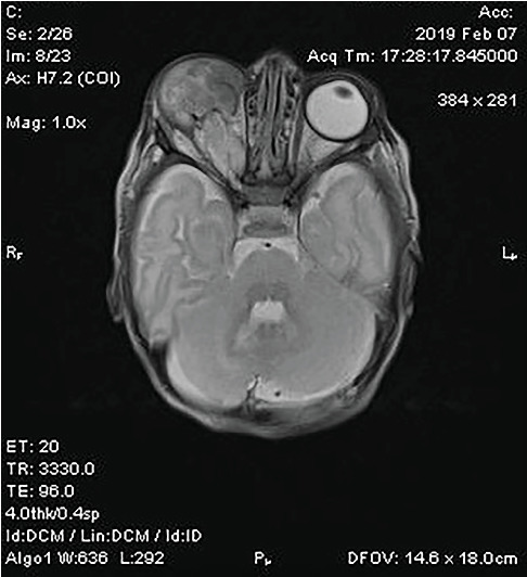
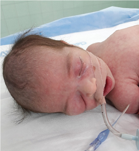
This case report describes a rare instance of primary orbital teratoma with anophthalmia in a neonate. A 6-day-old female presented with a congenital right orbital swelling and absence of visible ocular structures. MRI revealed a large, well-vascularized orbital mass without intracranial extension, accompanied by malformations in the right cerebral hemisphere. Histopathological examination confirmed a benign, mature/mixed teratoma comprising elements from all three germ layers, including neuroectoderm, mesoderm, and endoderm, with no evidence of malignancy. The patient underwent successful orbital exenteration with an implant at 3 weeks of age.

 求助内容:
求助内容: 应助结果提醒方式:


