András I. Försönits, Eszter Á. Tóth, Sára Jezsoviczky, Tünde Bárkai, Delaram Khamari, Alicia Galinsoga, Panna Királyhidi, Ágnes Kittel, Júlia Fazakas, Dorina Lenzinger, Hargita Hegyesi, Xabier Osteikoetxea, Tamás Visnovitz, Krisztina Pálóczi, Szilvia Bősze, Edit I. Buzás
{"title":"Improved Accessibility of Extracellular Vesicle Surface Molecules Upon Partial Removal of the Protein Corona by High Ionic Strength","authors":"András I. Försönits, Eszter Á. Tóth, Sára Jezsoviczky, Tünde Bárkai, Delaram Khamari, Alicia Galinsoga, Panna Királyhidi, Ágnes Kittel, Júlia Fazakas, Dorina Lenzinger, Hargita Hegyesi, Xabier Osteikoetxea, Tamás Visnovitz, Krisztina Pálóczi, Szilvia Bősze, Edit I. Buzás","doi":"10.1002/jev2.70124","DOIUrl":null,"url":null,"abstract":"<p>Recent studies have confirmed that a biomolecular corona forms around extracellular vesicles (EVs) in biofluids. However, there is limited data on how this adsorbed corona affects the accessibility of EV surface molecules. Here, we investigated various potential corona-stripping conditions for their ability to affect the immune detection of EVs. First, we artificially formed an EV corona around nascent HEK293T-PalmGFP cell-derived large EVs (lEVs) by incubating them with Cy5-labelled human plasma proteins. The co-localisation rate of plasma proteins and lEVs decreased significantly upon high-salt washing with NaCl, LiCl and KCl solutions, suggesting a considerable removal of the corona components. Additional evidence for corona modification was a significantly increased fluorescent annexin V binding to plasma lEVs and annexin V affinity capture of both THP1- and blood plasma-derived lEVs upon high-salt washing. A similar effect of high ionic strength was observed when THP1 lEVs were separated from a serum-containing medium, which allowed for corona formation, but not when EVs were produced under serum-free conditions. Using a MACSPlex kit and high-salt washing for small EVs from plasma and THP1 conditioned medium, we also demonstrated significantly improved immunodetection of 15 and 9 out of 37 surface markers, respectively. In this Technical Note, we present evidence that modifying the protein corona around EVs can significantly affect the immune detection of specific EV markers.</p>","PeriodicalId":15811,"journal":{"name":"Journal of Extracellular Vesicles","volume":"14 7","pages":""},"PeriodicalIF":14.5000,"publicationDate":"2025-07-14","publicationTypes":"Journal Article","fieldsOfStudy":null,"isOpenAccess":false,"openAccessPdf":"https://onlinelibrary.wiley.com/doi/epdf/10.1002/jev2.70124","citationCount":"0","resultStr":null,"platform":"Semanticscholar","paperid":null,"PeriodicalName":"Journal of Extracellular Vesicles","FirstCategoryId":"3","ListUrlMain":"https://isevjournals.onlinelibrary.wiley.com/doi/10.1002/jev2.70124","RegionNum":1,"RegionCategory":"医学","ArticlePicture":[],"TitleCN":null,"AbstractTextCN":null,"PMCID":null,"EPubDate":"","PubModel":"","JCR":"Q1","JCRName":"CELL BIOLOGY","Score":null,"Total":0}
引用次数: 0
Abstract
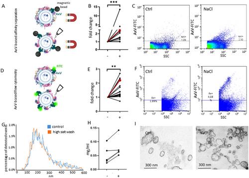
Recent studies have confirmed that a biomolecular corona forms around extracellular vesicles (EVs) in biofluids. However, there is limited data on how this adsorbed corona affects the accessibility of EV surface molecules. Here, we investigated various potential corona-stripping conditions for their ability to affect the immune detection of EVs. First, we artificially formed an EV corona around nascent HEK293T-PalmGFP cell-derived large EVs (lEVs) by incubating them with Cy5-labelled human plasma proteins. The co-localisation rate of plasma proteins and lEVs decreased significantly upon high-salt washing with NaCl, LiCl and KCl solutions, suggesting a considerable removal of the corona components. Additional evidence for corona modification was a significantly increased fluorescent annexin V binding to plasma lEVs and annexin V affinity capture of both THP1- and blood plasma-derived lEVs upon high-salt washing. A similar effect of high ionic strength was observed when THP1 lEVs were separated from a serum-containing medium, which allowed for corona formation, but not when EVs were produced under serum-free conditions. Using a MACSPlex kit and high-salt washing for small EVs from plasma and THP1 conditioned medium, we also demonstrated significantly improved immunodetection of 15 and 9 out of 37 surface markers, respectively. In this Technical Note, we present evidence that modifying the protein corona around EVs can significantly affect the immune detection of specific EV markers.
期刊介绍:
The Journal of Extracellular Vesicles is an open access research publication that focuses on extracellular vesicles, including microvesicles, exosomes, ectosomes, and apoptotic bodies. It serves as the official journal of the International Society for Extracellular Vesicles and aims to facilitate the exchange of data, ideas, and information pertaining to the chemistry, biology, and applications of extracellular vesicles. The journal covers various aspects such as the cellular and molecular mechanisms of extracellular vesicles biogenesis, technological advancements in their isolation, quantification, and characterization, the role and function of extracellular vesicles in biology, stem cell-derived extracellular vesicles and their biology, as well as the application of extracellular vesicles for pharmacological, immunological, or genetic therapies.
The Journal of Extracellular Vesicles is widely recognized and indexed by numerous services, including Biological Abstracts, BIOSIS Previews, Chemical Abstracts Service (CAS), Current Contents/Life Sciences, Directory of Open Access Journals (DOAJ), Journal Citation Reports/Science Edition, Google Scholar, ProQuest Natural Science Collection, ProQuest SciTech Collection, SciTech Premium Collection, PubMed Central/PubMed, Science Citation Index Expanded, ScienceOpen, and Scopus.


 求助内容:
求助内容: 应助结果提醒方式:


