{"title":"Calcium phosphate reinforced chitosan–carrageenan scaffolds: characterization and in vitro assessment for wound healing†","authors":"Vinita Patole, Gaurav Kavitkar, Ganesh Ingavle, Isha Behere, Ravindra Wavhale, Abhishek Jha, Sanjeevani Deshkar, Avinash Sanap and Pramod Sakpal","doi":"10.1039/D4PM00284A","DOIUrl":null,"url":null,"abstract":"<p >Wound healing is a multifaceted and dynamic biological process, which traditional wound dressings often fail to adequately support, leading to prolonged healing times. It would be highly beneficial to develop wound dressings with the ability to support biological processes such as cell proliferation and angiogenesis and deliver the active agents required to restore intracellular activities to promote wound healing. The current work aimed at developing a polyelectrolyte complex of chitosan (CH) and an anionic polymer, condensed with calcium phosphate (CaP) powder to attain antibacterial and angiogenic potential, cell proliferation, appropriate swelling index, and enhanced wound healing. Polyelectrolyte complexes (PECs) were formulated using chitosan (CH), as a cationic polymer and pectin (PE), sodium alginate (SA), and carrageenan (CA), respectively, as an anionic polymer through a lyophilization process. PEC formation was confirmed by FTIR, XRD, and DSC by observing the changes in their vibrational frequencies, structures, and thermal properties. SEM revealed the porous structure of the scaffolds. From the prepared PEC scaffolds, chitosan–carrageenan (CH-CA) was selected for further studies based on the swelling index, porosity, and degradation studies. Following the production of CaP powder using a microwave-assisted synthesis method, the powder was characterized by FTIR, SEM, XRD, and energy dispersive X-ray (EDX) techniques before being loaded onto CH-CA scaffolds. The results demonstrated approximately 60.75% release of calcium ions (Ca<small><sup>++</sup></small>) from the CH-CA scaffolds in PBS, pH 5.5, as analysed by atomic absorption spectroscopy (AAS) over 24 h. The scaffolds demonstrated a higher swelling index and exhibited antimicrobial activity against <em>E. coli</em> and <em>S. aureus</em>. The scaffolds were found to be hemocompatible and demonstrated angiogenic potential, evidenced by stimulating new blood vessel development in a chick yolk sac membrane assay. Cell proliferation studies demonstrated the cytocompatibility of the scaffolds, and improvement in the cell density of the L929 mouse fibroblast cell line was observed in a live/dead assay. In conclusion, the calcium-loaded CH-CA scaffolds demonstrated antimicrobial properties, increased angiogenesis, blood compatibility, and cell proliferation, indicating their potential as an appropriate wound dressing material.</p>","PeriodicalId":101141,"journal":{"name":"RSC Pharmaceutics","volume":" 4","pages":" 772-791"},"PeriodicalIF":0.0000,"publicationDate":"2025-05-05","publicationTypes":"Journal Article","fieldsOfStudy":null,"isOpenAccess":false,"openAccessPdf":"https://pubs.rsc.org/en/content/articlepdf/2025/pm/d4pm00284a?page=search","citationCount":"0","resultStr":null,"platform":"Semanticscholar","paperid":null,"PeriodicalName":"RSC Pharmaceutics","FirstCategoryId":"1085","ListUrlMain":"https://pubs.rsc.org/en/content/articlelanding/2025/pm/d4pm00284a","RegionNum":0,"RegionCategory":null,"ArticlePicture":[],"TitleCN":null,"AbstractTextCN":null,"PMCID":null,"EPubDate":"","PubModel":"","JCR":"","JCRName":"","Score":null,"Total":0}
引用次数: 0
Abstract
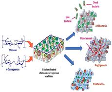
Wound healing is a multifaceted and dynamic biological process, which traditional wound dressings often fail to adequately support, leading to prolonged healing times. It would be highly beneficial to develop wound dressings with the ability to support biological processes such as cell proliferation and angiogenesis and deliver the active agents required to restore intracellular activities to promote wound healing. The current work aimed at developing a polyelectrolyte complex of chitosan (CH) and an anionic polymer, condensed with calcium phosphate (CaP) powder to attain antibacterial and angiogenic potential, cell proliferation, appropriate swelling index, and enhanced wound healing. Polyelectrolyte complexes (PECs) were formulated using chitosan (CH), as a cationic polymer and pectin (PE), sodium alginate (SA), and carrageenan (CA), respectively, as an anionic polymer through a lyophilization process. PEC formation was confirmed by FTIR, XRD, and DSC by observing the changes in their vibrational frequencies, structures, and thermal properties. SEM revealed the porous structure of the scaffolds. From the prepared PEC scaffolds, chitosan–carrageenan (CH-CA) was selected for further studies based on the swelling index, porosity, and degradation studies. Following the production of CaP powder using a microwave-assisted synthesis method, the powder was characterized by FTIR, SEM, XRD, and energy dispersive X-ray (EDX) techniques before being loaded onto CH-CA scaffolds. The results demonstrated approximately 60.75% release of calcium ions (Ca++) from the CH-CA scaffolds in PBS, pH 5.5, as analysed by atomic absorption spectroscopy (AAS) over 24 h. The scaffolds demonstrated a higher swelling index and exhibited antimicrobial activity against E. coli and S. aureus. The scaffolds were found to be hemocompatible and demonstrated angiogenic potential, evidenced by stimulating new blood vessel development in a chick yolk sac membrane assay. Cell proliferation studies demonstrated the cytocompatibility of the scaffolds, and improvement in the cell density of the L929 mouse fibroblast cell line was observed in a live/dead assay. In conclusion, the calcium-loaded CH-CA scaffolds demonstrated antimicrobial properties, increased angiogenesis, blood compatibility, and cell proliferation, indicating their potential as an appropriate wound dressing material.

 求助内容:
求助内容: 应助结果提醒方式:


