Surendra Reddy Gundam, Manasa Kethamreddy, Andy González Rivera, Aditya Bansal, Viktoria Krol, Daniella A. Sahagun, Joanna E. Kusmirek, Derek R. Johnson, Maliha Zahid, Val J. Lowe and Mukesh K. Pandey
{"title":"Synthesis and preliminary evaluation of cardiac imaging with [68Ga]Ga-NOTA-CTP in normal and infarcted CD1 mice†","authors":"Surendra Reddy Gundam, Manasa Kethamreddy, Andy González Rivera, Aditya Bansal, Viktoria Krol, Daniella A. Sahagun, Joanna E. Kusmirek, Derek R. Johnson, Maliha Zahid, Val J. Lowe and Mukesh K. Pandey","doi":"10.1039/D5PM00047E","DOIUrl":null,"url":null,"abstract":"<p >Cell-penetrating peptide-based probes for positron emission tomography (PET) are currently being developed for cardiac imaging. Herein, we have conjugated a synthetic 12 amino acids (NH<small><sub>2</sub></small>-APWHLSSQYSRT-COOH) cardiac targeting peptide (CTP) with a NOTA chelator for <small><sup>68</sup></small>Ga labeling. The [<small><sup>68</sup></small>Ga]Ga-NOTA-CTP was synthesized with a decay-corrected radiochemical yield of 68.9 ± 12.8% (<em>n</em> = 13) and molar activity (<em>A</em><small><sub>m</sub></small>) of 1.3 ± 0.5 GBq per μmol (<em>n</em> = 13). The tracer was evaluated in healthy and diseased CD1 mice with myocardial infarction following ligation of the left anterior descending artery. PET/CT imaging and <em>ex vivo</em> biodistribution revealed rapid (within 30 min) clearance of [<small><sup>68</sup></small>Ga]Ga-NOTA-CTP from the blood through renal and hepatobiliary excretion pathways in both healthy and infarcted animals. The uptake of [<small><sup>68</sup></small>Ga]Ga-NOTA-CTP in the heart of healthy and infarcted animals did not show any statistically significant difference for up to 120 min post-injection, but regional differences within healthy and infarcted hearts were detected with [<small><sup>68</sup></small>Ga]Ga-NOTA-CTP by PET/CT imaging at early time points post-injection. Within a healthy heart, the left ventricle standardized uptake value (SUV) was lower than the right ventricle SUV at 10–30 min post-injection. This regional difference between the left and right ventricles was absent in the infarcted heart, likely due to post-ligation changes.</p>","PeriodicalId":101141,"journal":{"name":"RSC Pharmaceutics","volume":" 4","pages":" 691-702"},"PeriodicalIF":0.0000,"publicationDate":"2025-05-29","publicationTypes":"Journal Article","fieldsOfStudy":null,"isOpenAccess":false,"openAccessPdf":"https://pubs.rsc.org/en/content/articlepdf/2025/pm/d5pm00047e?page=search","citationCount":"0","resultStr":null,"platform":"Semanticscholar","paperid":null,"PeriodicalName":"RSC Pharmaceutics","FirstCategoryId":"1085","ListUrlMain":"https://pubs.rsc.org/en/content/articlelanding/2025/pm/d5pm00047e","RegionNum":0,"RegionCategory":null,"ArticlePicture":[],"TitleCN":null,"AbstractTextCN":null,"PMCID":null,"EPubDate":"","PubModel":"","JCR":"","JCRName":"","Score":null,"Total":0}
引用次数: 0
Abstract
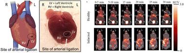
Cell-penetrating peptide-based probes for positron emission tomography (PET) are currently being developed for cardiac imaging. Herein, we have conjugated a synthetic 12 amino acids (NH2-APWHLSSQYSRT-COOH) cardiac targeting peptide (CTP) with a NOTA chelator for 68Ga labeling. The [68Ga]Ga-NOTA-CTP was synthesized with a decay-corrected radiochemical yield of 68.9 ± 12.8% (n = 13) and molar activity (Am) of 1.3 ± 0.5 GBq per μmol (n = 13). The tracer was evaluated in healthy and diseased CD1 mice with myocardial infarction following ligation of the left anterior descending artery. PET/CT imaging and ex vivo biodistribution revealed rapid (within 30 min) clearance of [68Ga]Ga-NOTA-CTP from the blood through renal and hepatobiliary excretion pathways in both healthy and infarcted animals. The uptake of [68Ga]Ga-NOTA-CTP in the heart of healthy and infarcted animals did not show any statistically significant difference for up to 120 min post-injection, but regional differences within healthy and infarcted hearts were detected with [68Ga]Ga-NOTA-CTP by PET/CT imaging at early time points post-injection. Within a healthy heart, the left ventricle standardized uptake value (SUV) was lower than the right ventricle SUV at 10–30 min post-injection. This regional difference between the left and right ventricles was absent in the infarcted heart, likely due to post-ligation changes.

 求助内容:
求助内容: 应助结果提醒方式:


