Marta Pietraszek, Marcin Stański, Joanna Marszał, Katarzyna Karmelita-Katulska, Anna Bartochowska, Andrzej Balcerowiak, Wojciech Gawęcki
{"title":"Detection of cholesteatoma recurrence by magnetic resonance imaging (DWI non-EPI sequence) - how can we minimise false results?","authors":"Marta Pietraszek, Marcin Stański, Joanna Marszał, Katarzyna Karmelita-Katulska, Anna Bartochowska, Andrzej Balcerowiak, Wojciech Gawęcki","doi":"10.5114/pjr/203991","DOIUrl":null,"url":null,"abstract":"<p><strong>Purpose: </strong>To evaluate the effectiveness of head magnetic resonance imaging (MRI) with the diffusion weighted imaging without echo-planar imaging (DWI non-EPI) sequence in detecting cholesteatoma recurrence, focusing on the analysis of false results.</p><p><strong>Material and methods: </strong>A retrospective study was conducted involving 156 patients diagnosed with cholesteatoma, who underwent reoperation between 2015 and 2021. All patients underwent preoperative MRI with the DWI non-EPI sequence. Data from surgical protocols, medical histories, outpatient records, and imaging results were analysed. MRI scans were reviewed by experienced radiologists and otosurgeons. The study was approved by the local Bioethics Committee.</p><p><strong>Results: </strong>Clinical and radiological concordance was found in 80% of patients. True positive results were observed in 77.5% of cases, while true negative results were noted in 2.5%. False positive results occurred in 8% of cases, mainly due to wax in the external auditory canal. False negative results were found in 12% of cases, often due to small or mural cholesteatomas. The sensitivity of MRI DWI non-EPI in detecting cholesteatoma was 87%.</p><p><strong>Conclusions: </strong>MRI DWI non-EPI is an effective tool for detecting cholesteatoma recurrence, potentially avoiding unnecessary second-look surgeries. Awareness of false positive and negative results is crucial, and correlation of MRI findings with clinical examinations is recommended. To minimise false results, ear cleaning before MRI and repeated examinations at intervals are advised.</p>","PeriodicalId":94174,"journal":{"name":"Polish journal of radiology","volume":"90 ","pages":"e318-e323"},"PeriodicalIF":0.0000,"publicationDate":"2025-06-25","publicationTypes":"Journal Article","fieldsOfStudy":null,"isOpenAccess":false,"openAccessPdf":"https://www.ncbi.nlm.nih.gov/pmc/articles/PMC12243516/pdf/","citationCount":"0","resultStr":null,"platform":"Semanticscholar","paperid":null,"PeriodicalName":"Polish journal of radiology","FirstCategoryId":"1085","ListUrlMain":"https://doi.org/10.5114/pjr/203991","RegionNum":0,"RegionCategory":null,"ArticlePicture":[],"TitleCN":null,"AbstractTextCN":null,"PMCID":null,"EPubDate":"2025/1/1 0:00:00","PubModel":"eCollection","JCR":"","JCRName":"","Score":null,"Total":0}
引用次数: 0
Abstract
Purpose: To evaluate the effectiveness of head magnetic resonance imaging (MRI) with the diffusion weighted imaging without echo-planar imaging (DWI non-EPI) sequence in detecting cholesteatoma recurrence, focusing on the analysis of false results.
Material and methods: A retrospective study was conducted involving 156 patients diagnosed with cholesteatoma, who underwent reoperation between 2015 and 2021. All patients underwent preoperative MRI with the DWI non-EPI sequence. Data from surgical protocols, medical histories, outpatient records, and imaging results were analysed. MRI scans were reviewed by experienced radiologists and otosurgeons. The study was approved by the local Bioethics Committee.
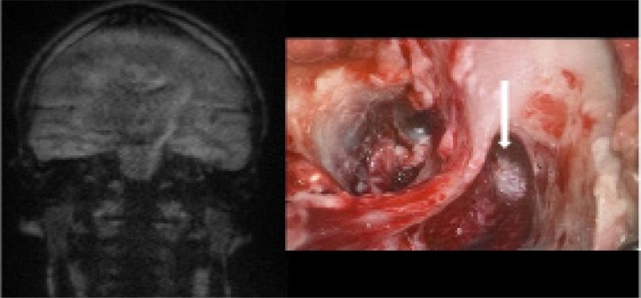
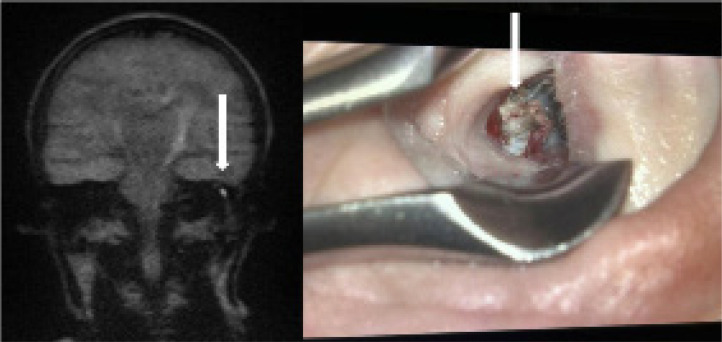
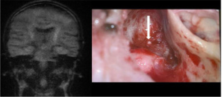
Results: Clinical and radiological concordance was found in 80% of patients. True positive results were observed in 77.5% of cases, while true negative results were noted in 2.5%. False positive results occurred in 8% of cases, mainly due to wax in the external auditory canal. False negative results were found in 12% of cases, often due to small or mural cholesteatomas. The sensitivity of MRI DWI non-EPI in detecting cholesteatoma was 87%.
Conclusions: MRI DWI non-EPI is an effective tool for detecting cholesteatoma recurrence, potentially avoiding unnecessary second-look surgeries. Awareness of false positive and negative results is crucial, and correlation of MRI findings with clinical examinations is recommended. To minimise false results, ear cleaning before MRI and repeated examinations at intervals are advised.

 求助内容:
求助内容: 应助结果提醒方式:


