Victor Matheus Mendonça de Araújo, Manuela Rodrigues Neiva Fernandes, Lívia Guerreiro de Barros Bentes, Rafael Silva Lemos, Herick Pampolha Huet de Bacelar, Luis Otávio Amaral Duarte Pinto
{"title":"\"Gotta have balls\": bovine testicles and video magnification imaging in the varicocelectomy training.","authors":"Victor Matheus Mendonça de Araújo, Manuela Rodrigues Neiva Fernandes, Lívia Guerreiro de Barros Bentes, Rafael Silva Lemos, Herick Pampolha Huet de Bacelar, Luis Otávio Amaral Duarte Pinto","doi":"10.1590/acb405225","DOIUrl":null,"url":null,"abstract":"<p><strong>Purpose: </strong>To describe the varicocelectomy model using bull testicles and to evaluate microsurgical practice using the surgical microscope and video magnification system.</p><p><strong>Methods: </strong>Bovine testicles and spermatic cords were used, with the medial portion of the cord left free for the microsurgical varicocelectomy technique. Twenty 3rd-year medical students were divided into two groups, the video magnification system group (VSMG) and the surgical microscope group (SMG), to simulate varicocelectomy in the proposed model. Five training sessions were carried out for both groups (D1, D7, D14, D21, and D28), as well as a reassessment (D63), with a checklist applied on the first, third, and fifth day of training and on reassessment.</p><p><strong>Results: </strong>The model provides practical support for training in instrument handling, dissection of structures, anatomical identification, and vessel ligation as an alternative to learning the surgical technique. There was a drop in training time as the weeks went by, with no significant difference between the groups. There was no statistical difference in time (D14, D28, D63) or scores between groups.</p><p><strong>Conclusion: </strong>Microsurgical varicocelectomy training using testicles and sperm cords of bovine origin in the video magnification system and surgical microscope contributed to the acquisition of skills.</p>","PeriodicalId":93850,"journal":{"name":"Acta cirurgica brasileira","volume":"40 ","pages":"e405225"},"PeriodicalIF":1.3000,"publicationDate":"2025-07-07","publicationTypes":"Journal Article","fieldsOfStudy":null,"isOpenAccess":false,"openAccessPdf":"https://www.ncbi.nlm.nih.gov/pmc/articles/PMC12233887/pdf/","citationCount":"0","resultStr":null,"platform":"Semanticscholar","paperid":null,"PeriodicalName":"Acta cirurgica brasileira","FirstCategoryId":"1085","ListUrlMain":"https://doi.org/10.1590/acb405225","RegionNum":0,"RegionCategory":null,"ArticlePicture":[],"TitleCN":null,"AbstractTextCN":null,"PMCID":null,"EPubDate":"2025/1/1 0:00:00","PubModel":"eCollection","JCR":"","JCRName":"","Score":null,"Total":0}
引用次数: 0
Abstract
Purpose: To describe the varicocelectomy model using bull testicles and to evaluate microsurgical practice using the surgical microscope and video magnification system.
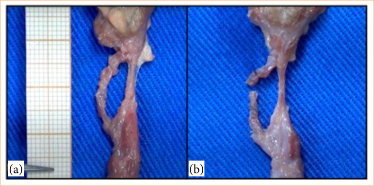
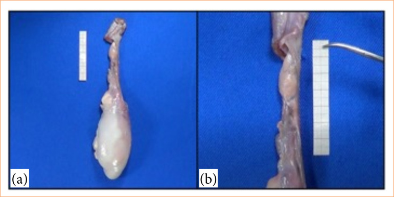
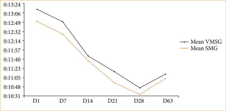
Methods: Bovine testicles and spermatic cords were used, with the medial portion of the cord left free for the microsurgical varicocelectomy technique. Twenty 3rd-year medical students were divided into two groups, the video magnification system group (VSMG) and the surgical microscope group (SMG), to simulate varicocelectomy in the proposed model. Five training sessions were carried out for both groups (D1, D7, D14, D21, and D28), as well as a reassessment (D63), with a checklist applied on the first, third, and fifth day of training and on reassessment.
Results: The model provides practical support for training in instrument handling, dissection of structures, anatomical identification, and vessel ligation as an alternative to learning the surgical technique. There was a drop in training time as the weeks went by, with no significant difference between the groups. There was no statistical difference in time (D14, D28, D63) or scores between groups.
Conclusion: Microsurgical varicocelectomy training using testicles and sperm cords of bovine origin in the video magnification system and surgical microscope contributed to the acquisition of skills.

 求助内容:
求助内容: 应助结果提醒方式:


