{"title":"Local anesthesia in upper eyelid surgery - Some considerations.","authors":"Samata Sharma, Jose M Ambat, Hirohiko Kakizaki","doi":"10.4103/sjopt.sjopt_59_25","DOIUrl":null,"url":null,"abstract":"<p><p>Local anesthesia injection into the upper eyelid is usually performed with the needle in the horizontal position, perpendicular to the vertically oriented subcutaneous vascular layer. This sometimes results in extensive eyelid ecchymosis and edema, causing prolonged downtime. The purpose of this article is to present an alternative way of infiltrating local anesthesia into the upper eyelids, taking into account the anatomic orientation of the subcutaneous vascular layer. The authors present how infiltration is done with the needle oriented parallel to the vertically oriented blood vessels underneath the skin. Inadvertent injury to the subcutaneous vascular layer was avoided with parallel alignment of the needle. After a certain volume of anesthesia injection into the central portion, it is extended with a glass bar or a cotton swab to infiltrate the whole eyelid. In case of insufficient infiltration to the medial or lateral edge, one more injection is added in the same manner. This reduced intraoperative bleeding, postoperative ecchymosis, and hematoma formation. When magnification is needed during surgery, placing a drop of clear liquid over the blood vessel gives an enlarged view and makes injection of local anesthesia easier. The authors recommend orienting the needle parallel to the vertical subcutaneous vascular layer with droplet magnification to minimize the chance of injury and bleeding and to decrease the incidence of postoperative swelling.</p>","PeriodicalId":46810,"journal":{"name":"Saudi Journal of Ophthalmology","volume":"39 2","pages":"141-142"},"PeriodicalIF":1.2000,"publicationDate":"2025-05-21","publicationTypes":"Journal Article","fieldsOfStudy":null,"isOpenAccess":false,"openAccessPdf":"https://www.ncbi.nlm.nih.gov/pmc/articles/PMC12240269/pdf/","citationCount":"0","resultStr":null,"platform":"Semanticscholar","paperid":null,"PeriodicalName":"Saudi Journal of Ophthalmology","FirstCategoryId":"1085","ListUrlMain":"https://doi.org/10.4103/sjopt.sjopt_59_25","RegionNum":0,"RegionCategory":null,"ArticlePicture":[],"TitleCN":null,"AbstractTextCN":null,"PMCID":null,"EPubDate":"2025/4/1 0:00:00","PubModel":"eCollection","JCR":"Q4","JCRName":"OPHTHALMOLOGY","Score":null,"Total":0}
引用次数: 0
Abstract
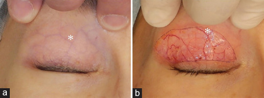
Local anesthesia injection into the upper eyelid is usually performed with the needle in the horizontal position, perpendicular to the vertically oriented subcutaneous vascular layer. This sometimes results in extensive eyelid ecchymosis and edema, causing prolonged downtime. The purpose of this article is to present an alternative way of infiltrating local anesthesia into the upper eyelids, taking into account the anatomic orientation of the subcutaneous vascular layer. The authors present how infiltration is done with the needle oriented parallel to the vertically oriented blood vessels underneath the skin. Inadvertent injury to the subcutaneous vascular layer was avoided with parallel alignment of the needle. After a certain volume of anesthesia injection into the central portion, it is extended with a glass bar or a cotton swab to infiltrate the whole eyelid. In case of insufficient infiltration to the medial or lateral edge, one more injection is added in the same manner. This reduced intraoperative bleeding, postoperative ecchymosis, and hematoma formation. When magnification is needed during surgery, placing a drop of clear liquid over the blood vessel gives an enlarged view and makes injection of local anesthesia easier. The authors recommend orienting the needle parallel to the vertical subcutaneous vascular layer with droplet magnification to minimize the chance of injury and bleeding and to decrease the incidence of postoperative swelling.
期刊介绍:
Saudi Journal of Ophthalmology is an English language, peer-reviewed scholarly publication in the area of ophthalmology. Saudi Journal of Ophthalmology publishes original papers, clinical studies, reviews and case reports. Saudi Journal of Ophthalmology is the official publication of the Saudi Ophthalmological Society and is published by King Saud University in collaboration with Elsevier and is edited by an international group of eminent researchers.

 求助内容:
求助内容: 应助结果提醒方式:


