Kan Ishijima, Jonnah K Teope, Yasuhiro Takahashi, Hirohiko Kakizaki
{"title":"Junction of lacrimal canaliculus to sac: Common or separated?","authors":"Kan Ishijima, Jonnah K Teope, Yasuhiro Takahashi, Hirohiko Kakizaki","doi":"10.4103/sjopt.sjopt_63_25","DOIUrl":null,"url":null,"abstract":"<p><strong>Purpose: </strong>This study aimed to determine whether the upper and lower canaliculi continue to the lacrimal sac through a common opening or separately.</p><p><strong>Methods: </strong>This retrospective cross-sectional study included the medical records of all patients who underwent external dacrycystorhinostomy or lacrimal intubation between April 1, 2024, and August 31, 2024. The canalicular opening into the lacrimal sac was examined by inserting a probe into the upper and lower puncta, and following its course into the sac using a microscope or canalicular endoscope.</p><p><strong>Results: </strong>Sixty-three lacrimal systems from 45 Japanese patients were included in the study. In 25 lacrimal systems (39.68%), the canaliculi shared a common opening into the lacrimal sac. In 38 lacrimal systems (60.32%), the upper and lower canaliculi opened separately into the lacrimal sac. One out of 18 bilateral cases (5.56%) showed a common opening on one side and separate openings on the other.</p><p><strong>Conclusion: </strong>Separate junction of the upper and lower canaliculi to the lacrimal sac was shown in 60.32% of the cases, a prevalence higher than previously reported. Both common and separated canalicular openings may be present in different sides of the same individual.</p>","PeriodicalId":46810,"journal":{"name":"Saudi Journal of Ophthalmology","volume":"39 2","pages":"155-157"},"PeriodicalIF":1.2000,"publicationDate":"2025-06-25","publicationTypes":"Journal Article","fieldsOfStudy":null,"isOpenAccess":false,"openAccessPdf":"https://www.ncbi.nlm.nih.gov/pmc/articles/PMC12240271/pdf/","citationCount":"0","resultStr":null,"platform":"Semanticscholar","paperid":null,"PeriodicalName":"Saudi Journal of Ophthalmology","FirstCategoryId":"1085","ListUrlMain":"https://doi.org/10.4103/sjopt.sjopt_63_25","RegionNum":0,"RegionCategory":null,"ArticlePicture":[],"TitleCN":null,"AbstractTextCN":null,"PMCID":null,"EPubDate":"2025/4/1 0:00:00","PubModel":"eCollection","JCR":"Q4","JCRName":"OPHTHALMOLOGY","Score":null,"Total":0}
引用次数: 0
Abstract
Purpose: This study aimed to determine whether the upper and lower canaliculi continue to the lacrimal sac through a common opening or separately.
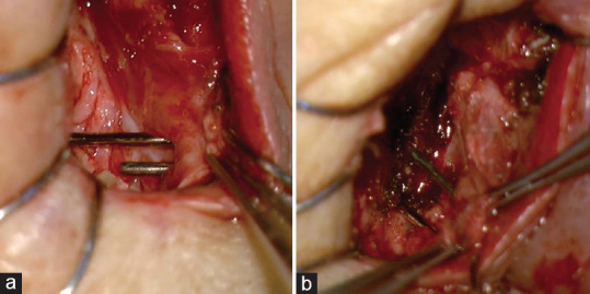
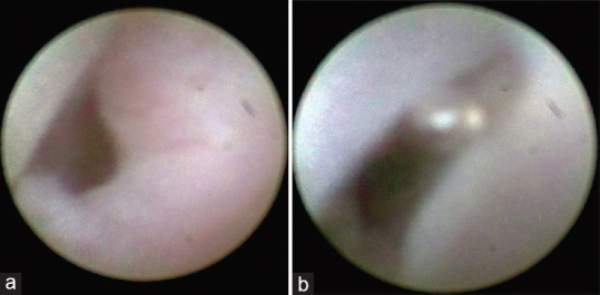
Methods: This retrospective cross-sectional study included the medical records of all patients who underwent external dacrycystorhinostomy or lacrimal intubation between April 1, 2024, and August 31, 2024. The canalicular opening into the lacrimal sac was examined by inserting a probe into the upper and lower puncta, and following its course into the sac using a microscope or canalicular endoscope.
Results: Sixty-three lacrimal systems from 45 Japanese patients were included in the study. In 25 lacrimal systems (39.68%), the canaliculi shared a common opening into the lacrimal sac. In 38 lacrimal systems (60.32%), the upper and lower canaliculi opened separately into the lacrimal sac. One out of 18 bilateral cases (5.56%) showed a common opening on one side and separate openings on the other.
Conclusion: Separate junction of the upper and lower canaliculi to the lacrimal sac was shown in 60.32% of the cases, a prevalence higher than previously reported. Both common and separated canalicular openings may be present in different sides of the same individual.
期刊介绍:
Saudi Journal of Ophthalmology is an English language, peer-reviewed scholarly publication in the area of ophthalmology. Saudi Journal of Ophthalmology publishes original papers, clinical studies, reviews and case reports. Saudi Journal of Ophthalmology is the official publication of the Saudi Ophthalmological Society and is published by King Saud University in collaboration with Elsevier and is edited by an international group of eminent researchers.

 求助内容:
求助内容: 应助结果提醒方式:


