African swine fever virus infection of porcine peripheral blood monocyte-derived macrophages induces the formation of tunneling nanotube-connected large vesicle-like cell segments: a potential mechanism for intercellular ASFV trafficking.
Brecht Droesbeke, Nadège Balmelle, Hans J Nauwynck, Herman Favoreel, Marylène Tignon
{"title":"African swine fever virus infection of porcine peripheral blood monocyte-derived macrophages induces the formation of tunneling nanotube-connected large vesicle-like cell segments: a potential mechanism for intercellular ASFV trafficking.","authors":"Brecht Droesbeke, Nadège Balmelle, Hans J Nauwynck, Herman Favoreel, Marylène Tignon","doi":"10.1186/s13567-025-01582-0","DOIUrl":null,"url":null,"abstract":"<p><p>African swine fever (ASF) is a highly fatal viral disease in pigs, with mortality rates that can reach 100%. The causative agent, African swine fever virus (ASFV), primarily targets cells of the mononuclear phagocytic system (MPS), particularly monocyte-derived macrophages (MDMs). Despite the severity of the disease, there are currently no effective antiviral treatments available in Europe. A significant barrier to therapeutic development is the limited understanding of how ASFV interacts with its primary target cells. A deeper understanding of the morphological changes induced by ASFV in infected cells is crucial to this effort. To address this knowledge gap, we used conventional and confocal immunofluorescence microscopy, as well as transmission electron microscopy, to investigate ASFV-infected primary MDMs. Our analysis revealed that ASFV infection leads to the formation of large cellular protrusions, which are characterized by vesicle-shaped cellular segments (CSs) at their tips. These protrusions contain all major cytoskeletal components, showing characteristics similar to those of tunneling nanotubes (TNTs). In 84.93% of the cases, the nucleus remained in the cell body (CB) near the viral factory. In the remaining cases, the nucleus was found within these CSs, whereas the viral factory was present in the CB. Additionally, 57.6% of the cells were in contact with the CS and distant cells, suggesting a potential mechanism for ASFV transmission. These findings suggest that ASFV induces cellular segmentation linked by TNT-like structures. Further research is needed to better understand the biogenesis and functional significance of these segmented cells, which could inform future strategies for combating ASFV.</p>","PeriodicalId":23658,"journal":{"name":"Veterinary Research","volume":"56 1","pages":"148"},"PeriodicalIF":3.5000,"publicationDate":"2025-07-10","publicationTypes":"Journal Article","fieldsOfStudy":null,"isOpenAccess":false,"openAccessPdf":"https://www.ncbi.nlm.nih.gov/pmc/articles/PMC12247315/pdf/","citationCount":"0","resultStr":null,"platform":"Semanticscholar","paperid":null,"PeriodicalName":"Veterinary Research","FirstCategoryId":"97","ListUrlMain":"https://doi.org/10.1186/s13567-025-01582-0","RegionNum":1,"RegionCategory":"农林科学","ArticlePicture":[],"TitleCN":null,"AbstractTextCN":null,"PMCID":null,"EPubDate":"","PubModel":"","JCR":"Q1","JCRName":"VETERINARY SCIENCES","Score":null,"Total":0}
引用次数: 0
Abstract
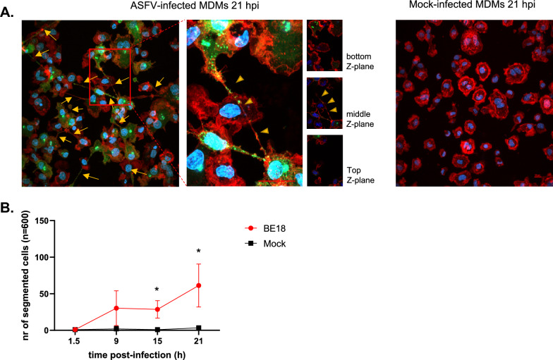
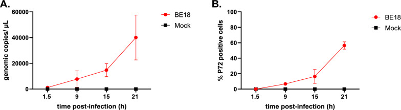
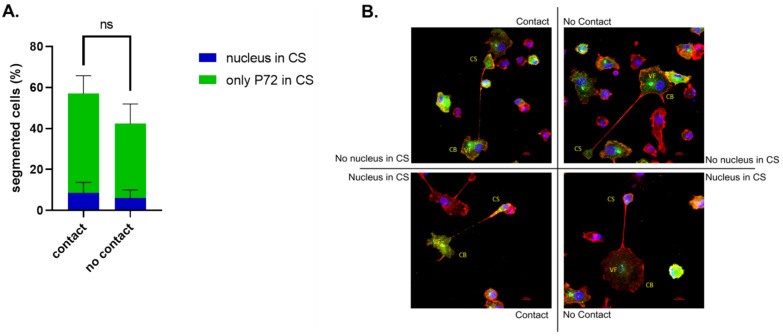
African swine fever (ASF) is a highly fatal viral disease in pigs, with mortality rates that can reach 100%. The causative agent, African swine fever virus (ASFV), primarily targets cells of the mononuclear phagocytic system (MPS), particularly monocyte-derived macrophages (MDMs). Despite the severity of the disease, there are currently no effective antiviral treatments available in Europe. A significant barrier to therapeutic development is the limited understanding of how ASFV interacts with its primary target cells. A deeper understanding of the morphological changes induced by ASFV in infected cells is crucial to this effort. To address this knowledge gap, we used conventional and confocal immunofluorescence microscopy, as well as transmission electron microscopy, to investigate ASFV-infected primary MDMs. Our analysis revealed that ASFV infection leads to the formation of large cellular protrusions, which are characterized by vesicle-shaped cellular segments (CSs) at their tips. These protrusions contain all major cytoskeletal components, showing characteristics similar to those of tunneling nanotubes (TNTs). In 84.93% of the cases, the nucleus remained in the cell body (CB) near the viral factory. In the remaining cases, the nucleus was found within these CSs, whereas the viral factory was present in the CB. Additionally, 57.6% of the cells were in contact with the CS and distant cells, suggesting a potential mechanism for ASFV transmission. These findings suggest that ASFV induces cellular segmentation linked by TNT-like structures. Further research is needed to better understand the biogenesis and functional significance of these segmented cells, which could inform future strategies for combating ASFV.
期刊介绍:
Veterinary Research is an open access journal that publishes high quality and novel research and review articles focusing on all aspects of infectious diseases and host-pathogen interaction in animals.
 求助内容:
求助内容: 应助结果提醒方式:


