Nilüfer Bıçakcı, Fatih Batı, Güler Silov, Banu Kırtıloğlu
{"title":"Incidental identification of elastofibroma dorsi in oncologic PET/CT imaging: a retrospective single-center analysis.","authors":"Nilüfer Bıçakcı, Fatih Batı, Güler Silov, Banu Kırtıloğlu","doi":"10.47717/turkjsurg.2025.2025-5-18","DOIUrl":null,"url":null,"abstract":"<p><strong>Objective: </strong>To evaluate the morphological and metabolic characteristics of incidentally detected elastofibroma dorsi (EFD) on F-18 florodeoksiglukoz (FDG) positron emission tomography/computed tomography (PET/CT) and their longitudinal changes in oncologic patients.</p><p><strong>Material and methods: </strong>We retrospectively reviewed 42 197 PET/CT scans performed at our institution between January 2019 and September 2023. EFD was incidentally identified in 20 patients (0.05%). Patient demographics, primary malignancy, lesion localization, dimensions, and maximum standardized uptake values (SUV<sub>max</sub>) were recorded. Measurements were obtained before treatment and at the next 3‑month follow‑up. Statistical analyses included Mann‑Whitney U, Shapiro-Wilk and Spearman correlation tests; significance was set at p<0.05.</p><p><strong>Results: </strong>The cohort comprised 17 females (85%) and 3 males (15%) with a median age of 67 years (range, 47-83). Primary diagnoses were breast cancer (n=8, 40%) and various other malignancies (n=12, 60%). Lesions were bilateral in 75% of cases. Pre‑treatment lesion size ranged from 10 to 55 mm; median SUV<sub>max</sub> was 2.4 (right) and 2.5 (left). No significant differences in baseline size or SUV<sub>max</sub> were observed between breast and other cancers. A moderate correlation existed between right and left SUV<sub>max</sub> (r=0.641; p=0.010). After 3 months, only the left longest diameter showed a statistically significant decrease (median, 45.0 mm vs. 43.0 mm; p=0.034), which may reflect measurement variability or positional factors rather than true biological change. SUV<sub>max</sub> values remained stable.</p><p><strong>Conclusion: </strong>Incidentally detected EFD on PET/CT exhibits low to moderate and stable FDG uptake and predominantly bilateral localization. Recognition of its characteristic features can prevent unnecessary interventions.</p>","PeriodicalId":23374,"journal":{"name":"Turkish Journal of Surgery","volume":" ","pages":"313-320"},"PeriodicalIF":0.6000,"publicationDate":"2025-09-03","publicationTypes":"Journal Article","fieldsOfStudy":null,"isOpenAccess":false,"openAccessPdf":"https://www.ncbi.nlm.nih.gov/pmc/articles/PMC12406630/pdf/","citationCount":"0","resultStr":null,"platform":"Semanticscholar","paperid":null,"PeriodicalName":"Turkish Journal of Surgery","FirstCategoryId":"1085","ListUrlMain":"https://doi.org/10.47717/turkjsurg.2025.2025-5-18","RegionNum":0,"RegionCategory":null,"ArticlePicture":[],"TitleCN":null,"AbstractTextCN":null,"PMCID":null,"EPubDate":"2025/7/11 0:00:00","PubModel":"Epub","JCR":"Q4","JCRName":"SURGERY","Score":null,"Total":0}
引用次数: 0
Abstract
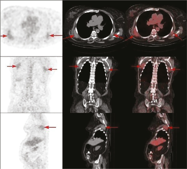
Objective: To evaluate the morphological and metabolic characteristics of incidentally detected elastofibroma dorsi (EFD) on F-18 florodeoksiglukoz (FDG) positron emission tomography/computed tomography (PET/CT) and their longitudinal changes in oncologic patients.
Material and methods: We retrospectively reviewed 42 197 PET/CT scans performed at our institution between January 2019 and September 2023. EFD was incidentally identified in 20 patients (0.05%). Patient demographics, primary malignancy, lesion localization, dimensions, and maximum standardized uptake values (SUVmax) were recorded. Measurements were obtained before treatment and at the next 3‑month follow‑up. Statistical analyses included Mann‑Whitney U, Shapiro-Wilk and Spearman correlation tests; significance was set at p<0.05.
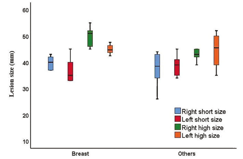
Results: The cohort comprised 17 females (85%) and 3 males (15%) with a median age of 67 years (range, 47-83). Primary diagnoses were breast cancer (n=8, 40%) and various other malignancies (n=12, 60%). Lesions were bilateral in 75% of cases. Pre‑treatment lesion size ranged from 10 to 55 mm; median SUVmax was 2.4 (right) and 2.5 (left). No significant differences in baseline size or SUVmax were observed between breast and other cancers. A moderate correlation existed between right and left SUVmax (r=0.641; p=0.010). After 3 months, only the left longest diameter showed a statistically significant decrease (median, 45.0 mm vs. 43.0 mm; p=0.034), which may reflect measurement variability or positional factors rather than true biological change. SUVmax values remained stable.
Conclusion: Incidentally detected EFD on PET/CT exhibits low to moderate and stable FDG uptake and predominantly bilateral localization. Recognition of its characteristic features can prevent unnecessary interventions.

 求助内容:
求助内容: 应助结果提醒方式:


