R Jablonowski, H B Andersson, C Fogarasi, H Engblom, H Arheden, P Kellman, M Carlsson, M Melin, I H Löfman, J Nickander, O Ö Braun
{"title":"Quantitative cardiovascular magnetic resonance myocardial perfusion can discriminate significant cardiac allograft vasculopathy: a multi-centre study.","authors":"R Jablonowski, H B Andersson, C Fogarasi, H Engblom, H Arheden, P Kellman, M Carlsson, M Melin, I H Löfman, J Nickander, O Ö Braun","doi":"10.1093/ehjci/jeaf201","DOIUrl":null,"url":null,"abstract":"<p><strong>Aims: </strong>Cardiac allograft vasculopathy (CAV) is a significant complication that contributes to both morbidity and mortality after heart transplantation. The aim of this study was to (i) assess if quantitative cardiovascular magnetic resonance (CMR) myocardial perfusion could detect different stages of CAV and (ii) establish a myocardial perfusion reserve (MPR) cut-off for significant CAV.</p><p><strong>Methods and results: </strong>Patients with a heart transplant who had performed a clinical CMR scan and invasive angiography at two centres in Sweden were included in the study (n = 110). Quantitative short-axis perfusion maps were acquired using single-bolus gadolinium contrast, dual-sequence perfusion imaging at rest and during stress. Global myocardial perfusion (MP) was averaged across all segments at rest and stress and MPR was defined as the ratio between stress and rest MP. All invasive angiographies were reported according to the International Heart and Lung Transplantation CAV classification. Patients were classified as follows: 53% (58/110) as CAV0, 38% (42/110) as CAV1, and 9% (10/110) as CAV2-3. There was a gradual decrease of stress MP and MPR with increased CAV grade. The MPR could discriminate CAV2-3 with an area under the curve-receiver operating characteristic of 0.88, 95% confidence interval 0.78-0.98, and using a cut-off of 2.2, the sensitivity was 100%, specificity was 68%, and positive and negative predictive values were 21 and 100%.</p><p><strong>Conclusion: </strong>In this multi-centre retrospective study, MPR assessed by CMR could discriminate CAV2-3 with both high sensitivity and negative predictive value and a cut-off of MPR 2.2 is suggested.</p>","PeriodicalId":12026,"journal":{"name":"European Heart Journal - Cardiovascular Imaging","volume":" ","pages":"1623-1630"},"PeriodicalIF":6.6000,"publicationDate":"2025-09-30","publicationTypes":"Journal Article","fieldsOfStudy":null,"isOpenAccess":false,"openAccessPdf":"https://www.ncbi.nlm.nih.gov/pmc/articles/PMC12481015/pdf/","citationCount":"0","resultStr":null,"platform":"Semanticscholar","paperid":null,"PeriodicalName":"European Heart Journal - Cardiovascular Imaging","FirstCategoryId":"3","ListUrlMain":"https://doi.org/10.1093/ehjci/jeaf201","RegionNum":1,"RegionCategory":"医学","ArticlePicture":[],"TitleCN":null,"AbstractTextCN":null,"PMCID":null,"EPubDate":"","PubModel":"","JCR":"Q1","JCRName":"CARDIAC & CARDIOVASCULAR SYSTEMS","Score":null,"Total":0}
引用次数: 0
Abstract
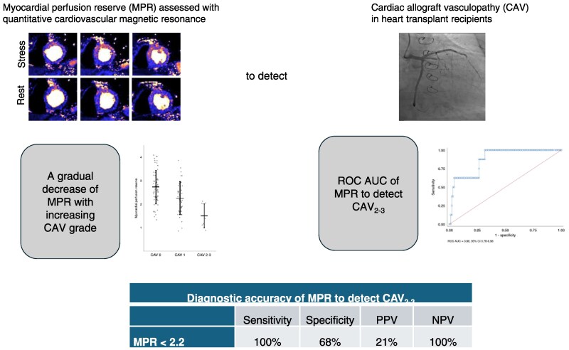
Aims: Cardiac allograft vasculopathy (CAV) is a significant complication that contributes to both morbidity and mortality after heart transplantation. The aim of this study was to (i) assess if quantitative cardiovascular magnetic resonance (CMR) myocardial perfusion could detect different stages of CAV and (ii) establish a myocardial perfusion reserve (MPR) cut-off for significant CAV.
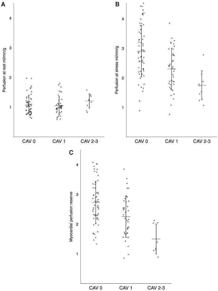
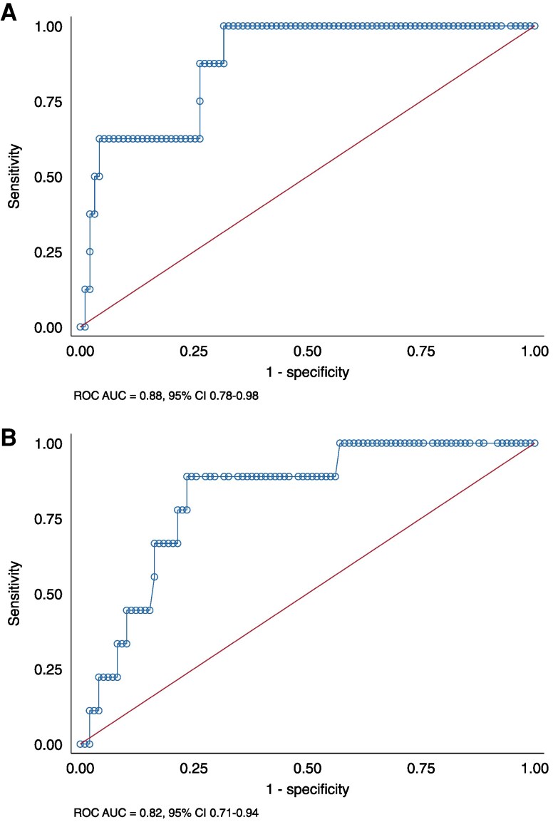
Methods and results: Patients with a heart transplant who had performed a clinical CMR scan and invasive angiography at two centres in Sweden were included in the study (n = 110). Quantitative short-axis perfusion maps were acquired using single-bolus gadolinium contrast, dual-sequence perfusion imaging at rest and during stress. Global myocardial perfusion (MP) was averaged across all segments at rest and stress and MPR was defined as the ratio between stress and rest MP. All invasive angiographies were reported according to the International Heart and Lung Transplantation CAV classification. Patients were classified as follows: 53% (58/110) as CAV0, 38% (42/110) as CAV1, and 9% (10/110) as CAV2-3. There was a gradual decrease of stress MP and MPR with increased CAV grade. The MPR could discriminate CAV2-3 with an area under the curve-receiver operating characteristic of 0.88, 95% confidence interval 0.78-0.98, and using a cut-off of 2.2, the sensitivity was 100%, specificity was 68%, and positive and negative predictive values were 21 and 100%.
Conclusion: In this multi-centre retrospective study, MPR assessed by CMR could discriminate CAV2-3 with both high sensitivity and negative predictive value and a cut-off of MPR 2.2 is suggested.
期刊介绍:
European Heart Journal – Cardiovascular Imaging is a monthly international peer reviewed journal dealing with Cardiovascular Imaging. It is an official publication of the European Association of Cardiovascular Imaging, a branch of the European Society of Cardiology.
The journal aims to publish the highest quality material, both scientific and clinical from all areas of cardiovascular imaging including echocardiography, magnetic resonance, computed tomography, nuclear and invasive imaging. A range of article types will be considered, including original research, reviews, editorials, image focus, letters and recommendation papers from relevant groups of the European Society of Cardiology. In addition it provides a forum for the exchange of information on all aspects of cardiovascular imaging.
 求助内容:
求助内容: 应助结果提醒方式:


