Thais Regina Dias Paini, Monique de Souza, Lidiane Vizioli de Castro-Hoshino, Isolde Terezinha Santos Previdelli, Antonio Carlos Bento, Francielle Sato, Fernanda Tsuzuki, Raquel Sano Suga Terada, Mauro Luciano Baesso, Renata Corrêa Pascotto
{"title":"Bleaching at acidic and basic pH for enamel and dentin whitening using internal and external techniques.","authors":"Thais Regina Dias Paini, Monique de Souza, Lidiane Vizioli de Castro-Hoshino, Isolde Terezinha Santos Previdelli, Antonio Carlos Bento, Francielle Sato, Fernanda Tsuzuki, Raquel Sano Suga Terada, Mauro Luciano Baesso, Renata Corrêa Pascotto","doi":"10.1590/1807-3107bor-2025.vol39.073","DOIUrl":null,"url":null,"abstract":"<p><p>This study aims to evaluate in vitro the whitening effects of gels containing hydrogen peroxide at acidic and basic pH using internal and external techniques. Forty artificially darkened bovine incisors were split into four groups: bleaching on the buccal surface with HPE (HP Blue 35%, FGM), at basic pH; PBE (Potenza Bianco 38%, PHS), at acidic pH; bleaching in the pulp chamber with HPI at basic pH; and with PBI at acidic pH. CIELab color parameters, microhardness, roughness, enamel demineralization (FT-Raman), and surface topography (SEM) were evaluated in enamel/dentin blocks after bleaching (T0) and seven days after immersion in saliva (T7). Bleaching agent diffusion was evaluated after 30 minutes, while pH was measured throughout the application period. Physicochemical analysis showed a reduction in the mineral-matrix ratio in both groups after bleaching and at 7 days. Roughness increased over time in the PBE and HPE groups. Porosity increased, but decreased after 7 days of immersion in saliva. Bleaching gels were different at T0 and T7, with acidic pH gel promoting greater whitening (T7) and enamel roughness. Raman scanning demonstrated that the bleaches propagated across the enamel layer with increased concentration at the dentin-enamel-junction and decreasing diffusion gradient along the dentin. The gel with acidic pH promoted a greater increase in enamel roughness and the 7-day immersion in saliva was not enough to restore initial roughness in either gel. The gel with basic pH showed a higher pH decay and a greater diffusion capacity in the dentin.</p>","PeriodicalId":9240,"journal":{"name":"Brazilian oral research","volume":"39 ","pages":"e073"},"PeriodicalIF":1.3000,"publicationDate":"2025-07-07","publicationTypes":"Journal Article","fieldsOfStudy":null,"isOpenAccess":false,"openAccessPdf":"https://www.ncbi.nlm.nih.gov/pmc/articles/PMC12237417/pdf/","citationCount":"0","resultStr":null,"platform":"Semanticscholar","paperid":null,"PeriodicalName":"Brazilian oral research","FirstCategoryId":"3","ListUrlMain":"https://doi.org/10.1590/1807-3107bor-2025.vol39.073","RegionNum":4,"RegionCategory":"医学","ArticlePicture":[],"TitleCN":null,"AbstractTextCN":null,"PMCID":null,"EPubDate":"2025/1/1 0:00:00","PubModel":"eCollection","JCR":"Q3","JCRName":"DENTISTRY, ORAL SURGERY & MEDICINE","Score":null,"Total":0}
引用次数: 0
Abstract
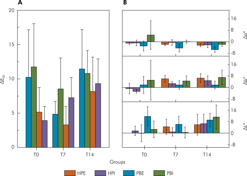
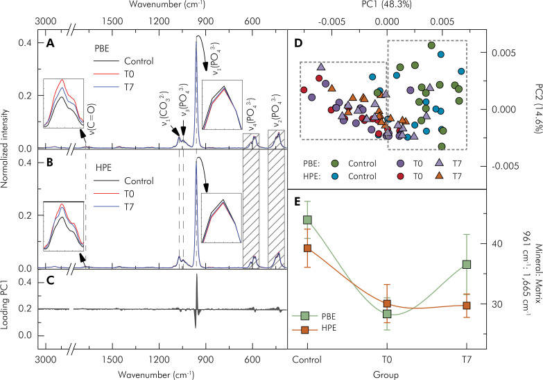
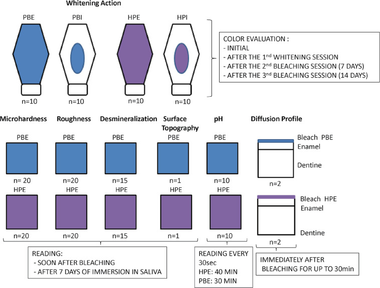
This study aims to evaluate in vitro the whitening effects of gels containing hydrogen peroxide at acidic and basic pH using internal and external techniques. Forty artificially darkened bovine incisors were split into four groups: bleaching on the buccal surface with HPE (HP Blue 35%, FGM), at basic pH; PBE (Potenza Bianco 38%, PHS), at acidic pH; bleaching in the pulp chamber with HPI at basic pH; and with PBI at acidic pH. CIELab color parameters, microhardness, roughness, enamel demineralization (FT-Raman), and surface topography (SEM) were evaluated in enamel/dentin blocks after bleaching (T0) and seven days after immersion in saliva (T7). Bleaching agent diffusion was evaluated after 30 minutes, while pH was measured throughout the application period. Physicochemical analysis showed a reduction in the mineral-matrix ratio in both groups after bleaching and at 7 days. Roughness increased over time in the PBE and HPE groups. Porosity increased, but decreased after 7 days of immersion in saliva. Bleaching gels were different at T0 and T7, with acidic pH gel promoting greater whitening (T7) and enamel roughness. Raman scanning demonstrated that the bleaches propagated across the enamel layer with increased concentration at the dentin-enamel-junction and decreasing diffusion gradient along the dentin. The gel with acidic pH promoted a greater increase in enamel roughness and the 7-day immersion in saliva was not enough to restore initial roughness in either gel. The gel with basic pH showed a higher pH decay and a greater diffusion capacity in the dentin.
 求助内容:
求助内容: 应助结果提醒方式:


