Clinical and Imaging Characteristics of Thalamic Infarction Combined With Moderate-to-Severe Stenosis of the Posterior Cerebral Artery: A Single-Center Experience.
Bailong Xin, Xiaomei Ye, Xiaoxue Liang, Yuzhen Wang, Yaozhuo Cai, Jingping Sun, Xueli Cai
{"title":"Clinical and Imaging Characteristics of Thalamic Infarction Combined With Moderate-to-Severe Stenosis of the Posterior Cerebral Artery: A Single-Center Experience.","authors":"Bailong Xin, Xiaomei Ye, Xiaoxue Liang, Yuzhen Wang, Yaozhuo Cai, Jingping Sun, Xueli Cai","doi":"10.1097/NRL.0000000000000634","DOIUrl":null,"url":null,"abstract":"<p><strong>Introduction: </strong>Ten patients with thalamic infarction caused by moderate-to-severe posterior cerebral artery (PCA) stenosis confirmed by computed tomography angiography (CTA) were enrolled. To better assess the vascular pathology, high-resolution magnetic resonance imaging (HRMRI) was subsequently used to evaluate the PCA in detail. In addition, we retrospectively analyzed clinical features, treatments, and prognostic outcomes.</p><p><strong>Case report: </strong>Ten patients were included, 7 males and 3 females, with an average age of 67.8±6.6 years. Past history includes: smoking (50%), drinking (30%), hypertension (70%), diabetes mellitus (40%), hyperlipidemia (10%), and cerebral infarction (10%). Clinical manifestations include sensory disorders (60%), motor disorders (50%), cognitive and consciousness disorders (10%), and language impairment (20%). HRMRI suggested that the PCA was moderately or severely stenosed in 4 cases, mildly stenosed in 5 cases, and normal in 1 case. It also suggested the presence of PCA atherosclerotic plaques in 9 patients.</p><p><strong>Conclusion: </strong>Antiplatelet therapy proves effective for this patient population. HRMRI identified atherosclerotic plaques mainly in the PCA's P1 and P2 segments. P1 stenosis often impairs consciousness, while P2 stenosis typically causes sensory/motor deficits. HRMRI aids in evaluating stenosis and plaque features for diagnosis and treatment guidance.</p>","PeriodicalId":49758,"journal":{"name":"Neurologist","volume":" ","pages":"293-298"},"PeriodicalIF":1.0000,"publicationDate":"2025-09-01","publicationTypes":"Journal Article","fieldsOfStudy":null,"isOpenAccess":false,"openAccessPdf":"https://www.ncbi.nlm.nih.gov/pmc/articles/PMC12404622/pdf/","citationCount":"0","resultStr":null,"platform":"Semanticscholar","paperid":null,"PeriodicalName":"Neurologist","FirstCategoryId":"3","ListUrlMain":"https://doi.org/10.1097/NRL.0000000000000634","RegionNum":4,"RegionCategory":"医学","ArticlePicture":[],"TitleCN":null,"AbstractTextCN":null,"PMCID":null,"EPubDate":"","PubModel":"","JCR":"Q4","JCRName":"CLINICAL NEUROLOGY","Score":null,"Total":0}
引用次数: 0
Abstract
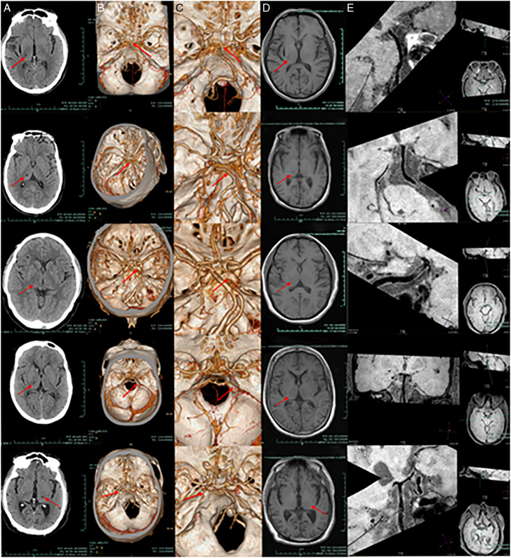
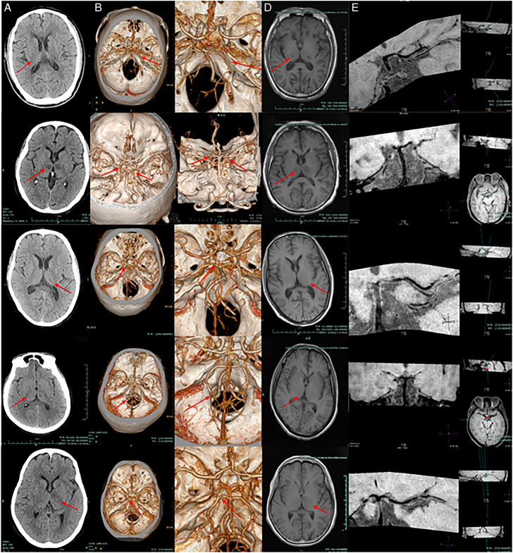
Introduction: Ten patients with thalamic infarction caused by moderate-to-severe posterior cerebral artery (PCA) stenosis confirmed by computed tomography angiography (CTA) were enrolled. To better assess the vascular pathology, high-resolution magnetic resonance imaging (HRMRI) was subsequently used to evaluate the PCA in detail. In addition, we retrospectively analyzed clinical features, treatments, and prognostic outcomes.
Case report: Ten patients were included, 7 males and 3 females, with an average age of 67.8±6.6 years. Past history includes: smoking (50%), drinking (30%), hypertension (70%), diabetes mellitus (40%), hyperlipidemia (10%), and cerebral infarction (10%). Clinical manifestations include sensory disorders (60%), motor disorders (50%), cognitive and consciousness disorders (10%), and language impairment (20%). HRMRI suggested that the PCA was moderately or severely stenosed in 4 cases, mildly stenosed in 5 cases, and normal in 1 case. It also suggested the presence of PCA atherosclerotic plaques in 9 patients.
Conclusion: Antiplatelet therapy proves effective for this patient population. HRMRI identified atherosclerotic plaques mainly in the PCA's P1 and P2 segments. P1 stenosis often impairs consciousness, while P2 stenosis typically causes sensory/motor deficits. HRMRI aids in evaluating stenosis and plaque features for diagnosis and treatment guidance.
期刊介绍:
The Neurologist publishes articles on topics of current interest to physicians treating patients with neurological diseases. The core of the journal is review articles focusing on clinically relevant issues. The journal also publishes case reports or case series which review the literature and put observations in perspective, as well as letters to the editor. Special features include the popular "10 Most Commonly Asked Questions" and the "Patient and Family Fact Sheet," a handy tear-out page that can be copied to hand out to patients and their caregivers.
 求助内容:
求助内容: 应助结果提醒方式:


