{"title":"PD-L1 expression on tumor cells and tumor-infiltrating immune cells in thymic epithelial tumors detected with SP142 and SP263 antibodies.","authors":"Kei Chubachi, Hisashi Tanaka, Kageaki Taima, Sadatomo Tasaka, Akira Kurose","doi":"10.1371/journal.pone.0327792","DOIUrl":null,"url":null,"abstract":"<p><strong>Introduction: </strong>Programmed death-ligand 1 (PD-L1) expression in various tumors is known to correlate with the efficacy of immune checkpoint inhibitors; however, evaluation of PD-L1 expression in thymic epithelial tumors (TETs) using multiple antibodies are limited. We retrospectivity evaluated PD-L1 expression in thymomas and thymic carcinomas using two antibodies, SP142 and SP263, and compared their expression rates in each type of TETs.</p><p><strong>Materials and methods: </strong>We retrospectively included 37 cases of thymoma and 11 cases of thymic carcinoma that were histologically diagnosed between January 2000 and December 2020. PD-L1 expression was assessed using SP142 and SP263 antibodies with semi-quantitative scoring.</p><p><strong>Results: </strong>The concordance rate for PD-L1 positivity between SP142 and SP263 was 81.2%, whereas the concordance rate for high PD-L1 expression was 83.3%. SP142 showed positive PD-L1 expression in 23 (62%) thymoma cases and eight (73%) thymic carcinoma cases. In contrast, SP263 antibody showed positive PD-L1 expression in 31 (84%) cases of thymoma and 9 (82%) cases of thymic carcinoma. In addition, type B thymomas exhibited significantly higher PD-L1 positivity than other thymoma types. The tumor-infiltrating lymphocytes were mostly CD3 and CD8 positive. No significant difference in overall survival was observed between the high and low PD-L1 expression groups in thymic carcinoma.</p><p><strong>Conclusion: </strong>PD-L1 expression rate was high in TETs, with variations depending on the antibody used and histological subtype. SP263 showed higher PD-L1 expression compared to SP142. The type of the antibody used should be considered when evaluating PD-L1 expression in TETs.</p>","PeriodicalId":20189,"journal":{"name":"PLoS ONE","volume":"20 7","pages":"e0327792"},"PeriodicalIF":2.6000,"publicationDate":"2025-07-09","publicationTypes":"Journal Article","fieldsOfStudy":null,"isOpenAccess":false,"openAccessPdf":"https://www.ncbi.nlm.nih.gov/pmc/articles/PMC12240378/pdf/","citationCount":"0","resultStr":null,"platform":"Semanticscholar","paperid":null,"PeriodicalName":"PLoS ONE","FirstCategoryId":"103","ListUrlMain":"https://doi.org/10.1371/journal.pone.0327792","RegionNum":3,"RegionCategory":"综合性期刊","ArticlePicture":[],"TitleCN":null,"AbstractTextCN":null,"PMCID":null,"EPubDate":"2025/1/1 0:00:00","PubModel":"eCollection","JCR":"Q1","JCRName":"MULTIDISCIPLINARY SCIENCES","Score":null,"Total":0}
引用次数: 0
Abstract
Introduction: Programmed death-ligand 1 (PD-L1) expression in various tumors is known to correlate with the efficacy of immune checkpoint inhibitors; however, evaluation of PD-L1 expression in thymic epithelial tumors (TETs) using multiple antibodies are limited. We retrospectivity evaluated PD-L1 expression in thymomas and thymic carcinomas using two antibodies, SP142 and SP263, and compared their expression rates in each type of TETs.
Materials and methods: We retrospectively included 37 cases of thymoma and 11 cases of thymic carcinoma that were histologically diagnosed between January 2000 and December 2020. PD-L1 expression was assessed using SP142 and SP263 antibodies with semi-quantitative scoring.
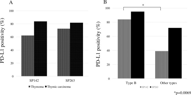
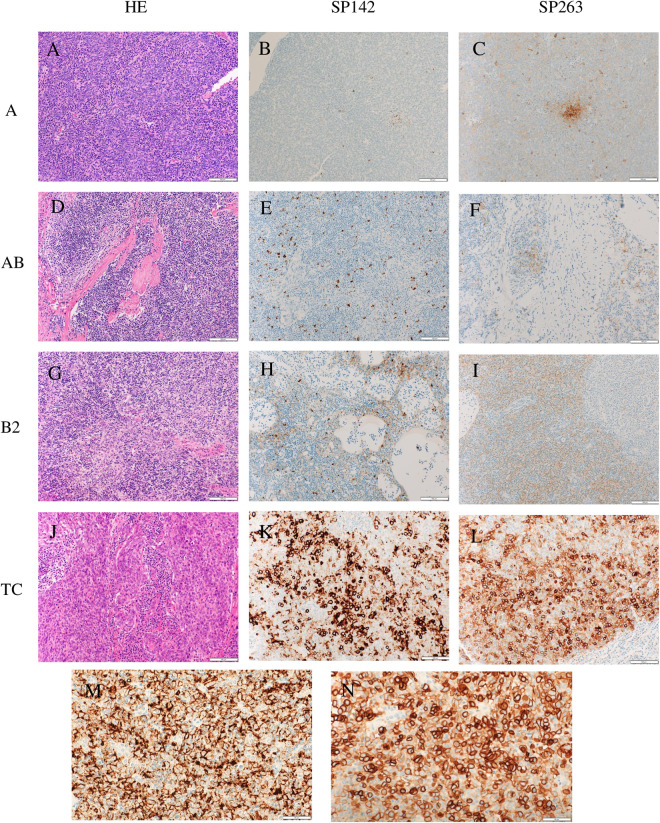
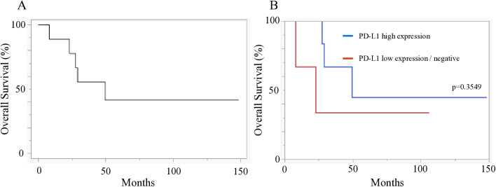
Results: The concordance rate for PD-L1 positivity between SP142 and SP263 was 81.2%, whereas the concordance rate for high PD-L1 expression was 83.3%. SP142 showed positive PD-L1 expression in 23 (62%) thymoma cases and eight (73%) thymic carcinoma cases. In contrast, SP263 antibody showed positive PD-L1 expression in 31 (84%) cases of thymoma and 9 (82%) cases of thymic carcinoma. In addition, type B thymomas exhibited significantly higher PD-L1 positivity than other thymoma types. The tumor-infiltrating lymphocytes were mostly CD3 and CD8 positive. No significant difference in overall survival was observed between the high and low PD-L1 expression groups in thymic carcinoma.
Conclusion: PD-L1 expression rate was high in TETs, with variations depending on the antibody used and histological subtype. SP263 showed higher PD-L1 expression compared to SP142. The type of the antibody used should be considered when evaluating PD-L1 expression in TETs.
期刊介绍:
PLOS ONE is an international, peer-reviewed, open-access, online publication. PLOS ONE welcomes reports on primary research from any scientific discipline. It provides:
* Open-access—freely accessible online, authors retain copyright
* Fast publication times
* Peer review by expert, practicing researchers
* Post-publication tools to indicate quality and impact
* Community-based dialogue on articles
* Worldwide media coverage

 求助内容:
求助内容: 应助结果提醒方式:


