Wentao Fu, Kun Liu, Yan Zhang, Jing Wang, Wan Cai, Yaoyao Wu, Hao Chi, Wen Ge
{"title":"Cardiac calcified amorphous tumor in the right atrium: a rare cardiac neoplasm.","authors":"Wentao Fu, Kun Liu, Yan Zhang, Jing Wang, Wan Cai, Yaoyao Wu, Hao Chi, Wen Ge","doi":"10.1186/s13019-025-03501-y","DOIUrl":null,"url":null,"abstract":"<p><strong>Background: </strong>Cardiac calcified amorphous tumors (CATs) represent rare, nonneoplastic intraluminal heart masses, with limited case reports in existing literature. Asymptomatic cases localized in the right atrium are particularly unusual.</p><p><strong>Case presentation: </strong>An asymptomatic 46-year-old male was discovered to have a cardiac mass upon echocardiograph. Echocardiography revealed a 13.2 × 11.8 mm pedunculated mass in the right atrium, attached to the interatrial septum. Then we performed surgical treatment. Histopathology revealed some myocardial tissue, a powdery stained, calcified amorphous area, and a few localized lymphocytes and red blood cells. The final diagnosis confirmed a cardiac CAT.</p><p><strong>Conclusions: </strong>CATs, rarely occurring endocardium-based pseudotumors, comprise calcium nodules and amorphous fibrin material. Typically presenting as a calcified pedunculated mass, they may arise in any heart chamber, with a significant propensity for distal embolism. Differentiating CATs from calcified atrial myxomas, calcified thrombi, or other cardiac tumors is challenging. Histopathology remains a critical diagnostic cornerstone. Although complete surgical resection is the recommended treatment, anticoagulation and ongoing surveillance may serve as viable alternatives when primary treatment, surgical resection, is deemed excessively hazardous.</p>","PeriodicalId":15201,"journal":{"name":"Journal of Cardiothoracic Surgery","volume":"20 1","pages":"288"},"PeriodicalIF":1.5000,"publicationDate":"2025-07-09","publicationTypes":"Journal Article","fieldsOfStudy":null,"isOpenAccess":false,"openAccessPdf":"https://www.ncbi.nlm.nih.gov/pmc/articles/PMC12239440/pdf/","citationCount":"0","resultStr":null,"platform":"Semanticscholar","paperid":null,"PeriodicalName":"Journal of Cardiothoracic Surgery","FirstCategoryId":"3","ListUrlMain":"https://doi.org/10.1186/s13019-025-03501-y","RegionNum":4,"RegionCategory":"医学","ArticlePicture":[],"TitleCN":null,"AbstractTextCN":null,"PMCID":null,"EPubDate":"","PubModel":"","JCR":"Q3","JCRName":"CARDIAC & CARDIOVASCULAR SYSTEMS","Score":null,"Total":0}
引用次数: 0
Abstract
Background: Cardiac calcified amorphous tumors (CATs) represent rare, nonneoplastic intraluminal heart masses, with limited case reports in existing literature. Asymptomatic cases localized in the right atrium are particularly unusual.
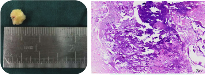
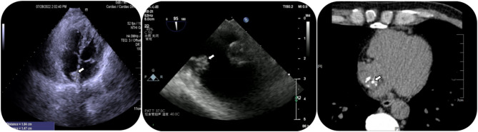
Case presentation: An asymptomatic 46-year-old male was discovered to have a cardiac mass upon echocardiograph. Echocardiography revealed a 13.2 × 11.8 mm pedunculated mass in the right atrium, attached to the interatrial septum. Then we performed surgical treatment. Histopathology revealed some myocardial tissue, a powdery stained, calcified amorphous area, and a few localized lymphocytes and red blood cells. The final diagnosis confirmed a cardiac CAT.
Conclusions: CATs, rarely occurring endocardium-based pseudotumors, comprise calcium nodules and amorphous fibrin material. Typically presenting as a calcified pedunculated mass, they may arise in any heart chamber, with a significant propensity for distal embolism. Differentiating CATs from calcified atrial myxomas, calcified thrombi, or other cardiac tumors is challenging. Histopathology remains a critical diagnostic cornerstone. Although complete surgical resection is the recommended treatment, anticoagulation and ongoing surveillance may serve as viable alternatives when primary treatment, surgical resection, is deemed excessively hazardous.
期刊介绍:
Journal of Cardiothoracic Surgery is an open access journal that encompasses all aspects of research in the field of Cardiology, and Cardiothoracic and Vascular Surgery. The journal publishes original scientific research documenting clinical and experimental advances in cardiac, vascular and thoracic surgery, and related fields.
Topics of interest include surgical techniques, survival rates, surgical complications and their outcomes; along with basic sciences, pediatric conditions, transplantations and clinical trials.
Journal of Cardiothoracic Surgery is of interest to cardiothoracic and vascular surgeons, cardiothoracic anaesthesiologists, cardiologists, chest physicians, and allied health professionals.
 求助内容:
求助内容: 应助结果提醒方式:


