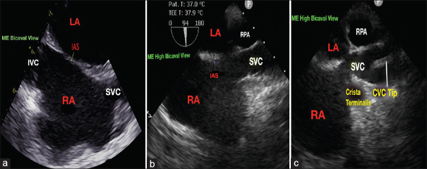
Superior vena cava and right pulmonary artery junction - An intraoperative transesophageal echocardiographic landmark for central venous catheter tip position: A prospective observational study.
A Jagadish, Saravana Babu, Subin Sukesan, Prasanta K Dash, Shrinivas V Gadhinglajkar, Bineesh K Radhakrishnan
{"title":"Superior vena cava and right pulmonary artery junction - An intraoperative transesophageal echocardiographic landmark for central venous catheter tip position: A prospective observational study.","authors":"A Jagadish, Saravana Babu, Subin Sukesan, Prasanta K Dash, Shrinivas V Gadhinglajkar, Bineesh K Radhakrishnan","doi":"10.4103/joacp.joacp_103_24","DOIUrl":null,"url":null,"abstract":"<p><strong>Background and aims: </strong>The main aim of this study was to evaluate the efficacy of real-time intraoperative transesophageal echocardiography (TEE) in guiding the central venous catheter (CVC) tip placement at the superior vena cava (SVC)-right pulmonary artery (RPA) junction.</p><p><strong>Materials and methods: </strong>One hundred patients aged between 18 and 65 years undergoing elective cardiac surgery were enrolled in the study. In the operation room, under ultrasound guidance, right internal jugular vein was punctured and CVC was inserted. The primary outcome was to determine the accuracy of placing the CVC tip under TEE guidance at the SVC-RPA junction by intraoperative surgical palpation and to correlate between the preoperative chest radiograph-predicted CVC depth and the TEE-guided placement of CVC depth. The secondary outcomes were to evaluate the position of CVC tip in relation to the carina in postoperative chest radiograph and the incidence of complications.</p><p><strong>Results: </strong>A total of 98 patients were included in the analysis. The CVC tip was palpable by the surgeon intraoperatively at the SVC-RPA junction in 76 patients (77.6%). A significant direct correlation was observed between the predicted preoperative CVC depth and TEE-guided placement of CVC depth (<i>r</i> = 0.7441, <i>P</i> < 0.0001). In the postoperative chest radiograph, 78 (79.5%) patients had the CVC tip positioned above the carina. Twenty-nine patients had atrial ectopics and six patients had ventricular ectopics during CVC insertion.</p><p><strong>Conclusions: </strong>TEE-guided SVC-RPA junction is an accurate landmark for the intraoperative positioning of CVC tip in the extra-pericardial portion of SVC to prevent life-threatening cardiac complications.</p>","PeriodicalId":14946,"journal":{"name":"Journal of Anaesthesiology, Clinical Pharmacology","volume":"41 3","pages":"427-432"},"PeriodicalIF":1.1000,"publicationDate":"2025-07-01","publicationTypes":"Journal Article","fieldsOfStudy":null,"isOpenAccess":false,"openAccessPdf":"https://www.ncbi.nlm.nih.gov/pmc/articles/PMC12237190/pdf/","citationCount":"0","resultStr":null,"platform":"Semanticscholar","paperid":null,"PeriodicalName":"Journal of Anaesthesiology, Clinical Pharmacology","FirstCategoryId":"1085","ListUrlMain":"https://doi.org/10.4103/joacp.joacp_103_24","RegionNum":0,"RegionCategory":null,"ArticlePicture":[],"TitleCN":null,"AbstractTextCN":null,"PMCID":null,"EPubDate":"2025/6/19 0:00:00","PubModel":"Epub","JCR":"Q3","JCRName":"PHARMACOLOGY & PHARMACY","Score":null,"Total":0}
引用次数: 0
Abstract
Background and aims: The main aim of this study was to evaluate the efficacy of real-time intraoperative transesophageal echocardiography (TEE) in guiding the central venous catheter (CVC) tip placement at the superior vena cava (SVC)-right pulmonary artery (RPA) junction.
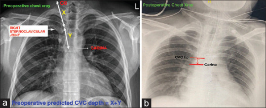
Materials and methods: One hundred patients aged between 18 and 65 years undergoing elective cardiac surgery were enrolled in the study. In the operation room, under ultrasound guidance, right internal jugular vein was punctured and CVC was inserted. The primary outcome was to determine the accuracy of placing the CVC tip under TEE guidance at the SVC-RPA junction by intraoperative surgical palpation and to correlate between the preoperative chest radiograph-predicted CVC depth and the TEE-guided placement of CVC depth. The secondary outcomes were to evaluate the position of CVC tip in relation to the carina in postoperative chest radiograph and the incidence of complications.
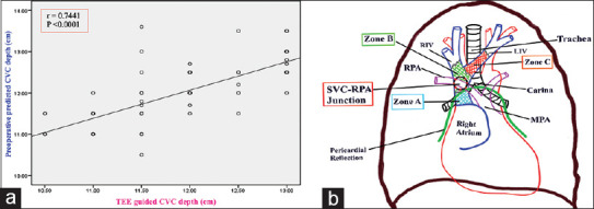
Results: A total of 98 patients were included in the analysis. The CVC tip was palpable by the surgeon intraoperatively at the SVC-RPA junction in 76 patients (77.6%). A significant direct correlation was observed between the predicted preoperative CVC depth and TEE-guided placement of CVC depth (r = 0.7441, P < 0.0001). In the postoperative chest radiograph, 78 (79.5%) patients had the CVC tip positioned above the carina. Twenty-nine patients had atrial ectopics and six patients had ventricular ectopics during CVC insertion.
Conclusions: TEE-guided SVC-RPA junction is an accurate landmark for the intraoperative positioning of CVC tip in the extra-pericardial portion of SVC to prevent life-threatening cardiac complications.
期刊介绍:
The JOACP publishes original peer-reviewed research and clinical work in all branches of anaesthesiology, pain, critical care and perioperative medicine including the application to basic sciences. In addition, the journal publishes review articles, special articles, brief communications/reports, case reports, and reports of new equipment, letters to editor, book reviews and obituaries. It is international in scope and comprehensive in coverage.

 求助内容:
求助内容: 应助结果提醒方式:


