{"title":"Assessment of condylar and glenoid fossa dimension in various sagittal skeletal malocclusions.","authors":"Ashish Ravi Kallur, Remmiya Mary Varghese","doi":"10.4103/jos.jos_105_24","DOIUrl":null,"url":null,"abstract":"<p><strong>Introduction: </strong>Jaw mobility and oral function depend on the temporomandibular joint (TMJ), with the dimensions of the mandibular condyle and glenoid fossa being important for understanding dental and orthodontic conditions. Additionally, advanced imaging, like cone-beam computed tomography (CBCT), has improved the study of condylar morphology, aiding diagnosis and treatment planning.</p><p><strong>Aim: </strong>To perform morphometric analysis of the TMJs using CBCT in all three dimensions in various skeletal malocclusions.</p><p><strong>Materials and methods: </strong>CBCT images of 84 patients were collected for the study. FACAD (ILEXIS AB, Linköping, Sweden) software was used to divide the patients into Class I, II, and III skeletal malocclusions. Dolphin software was used to measure the width, length, and height of condyles and glenoid fossa of the patients from the CBCT images. The Shapiro-Wilk test was used to check normality. ANOVA test was performed to assess the statistical significance of the results between the 3 groups.</p><p><strong>Results: </strong>The sample consisted of 30 patients with class I skeletal relation, 34 patients with class II relation, and 20 patients with class III relation. In class I relation, the average condylar height is found to be 16.32 mm ± 2.16 mm, width is 16.47 mm ± 2.61 mm, and length 7.65 mm ± 1.5 mm. The average dimensions of the glenoid fossa in class I skeletal relation were measured to be 19.93 mm ± 2.64 mm in width, 13.93 mm ± 1.45 mm in length, and 6.4 mm ± 1.49 mm in height.</p><p><strong>Conclusion: </strong>A statistically significant difference was noted in the condylar width and condylar height among the various skeletal malocclusions. The morphological parameters assessed in this study require more investigation to fully understand the mechanisms underlying them and investigate the consequences for orthodontic treatment and TMJ health.</p>","PeriodicalId":16604,"journal":{"name":"Journal of Orthodontic Science","volume":"14 ","pages":"16"},"PeriodicalIF":0.0000,"publicationDate":"2025-06-10","publicationTypes":"Journal Article","fieldsOfStudy":null,"isOpenAccess":false,"openAccessPdf":"https://www.ncbi.nlm.nih.gov/pmc/articles/PMC12237002/pdf/","citationCount":"0","resultStr":null,"platform":"Semanticscholar","paperid":null,"PeriodicalName":"Journal of Orthodontic Science","FirstCategoryId":"1085","ListUrlMain":"https://doi.org/10.4103/jos.jos_105_24","RegionNum":0,"RegionCategory":null,"ArticlePicture":[],"TitleCN":null,"AbstractTextCN":null,"PMCID":null,"EPubDate":"2025/1/1 0:00:00","PubModel":"eCollection","JCR":"Q2","JCRName":"Dentistry","Score":null,"Total":0}
引用次数: 0
Abstract
Introduction: Jaw mobility and oral function depend on the temporomandibular joint (TMJ), with the dimensions of the mandibular condyle and glenoid fossa being important for understanding dental and orthodontic conditions. Additionally, advanced imaging, like cone-beam computed tomography (CBCT), has improved the study of condylar morphology, aiding diagnosis and treatment planning.
Aim: To perform morphometric analysis of the TMJs using CBCT in all three dimensions in various skeletal malocclusions.
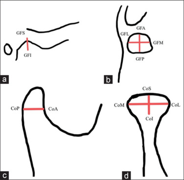
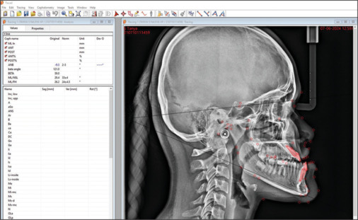
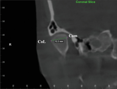
Materials and methods: CBCT images of 84 patients were collected for the study. FACAD (ILEXIS AB, Linköping, Sweden) software was used to divide the patients into Class I, II, and III skeletal malocclusions. Dolphin software was used to measure the width, length, and height of condyles and glenoid fossa of the patients from the CBCT images. The Shapiro-Wilk test was used to check normality. ANOVA test was performed to assess the statistical significance of the results between the 3 groups.
Results: The sample consisted of 30 patients with class I skeletal relation, 34 patients with class II relation, and 20 patients with class III relation. In class I relation, the average condylar height is found to be 16.32 mm ± 2.16 mm, width is 16.47 mm ± 2.61 mm, and length 7.65 mm ± 1.5 mm. The average dimensions of the glenoid fossa in class I skeletal relation were measured to be 19.93 mm ± 2.64 mm in width, 13.93 mm ± 1.45 mm in length, and 6.4 mm ± 1.49 mm in height.
Conclusion: A statistically significant difference was noted in the condylar width and condylar height among the various skeletal malocclusions. The morphological parameters assessed in this study require more investigation to fully understand the mechanisms underlying them and investigate the consequences for orthodontic treatment and TMJ health.

 求助内容:
求助内容: 应助结果提醒方式:


