{"title":"α-Synuclein-Assembled Synaptic Vesicle Pools at the Presynaptic Terminal: A Study of α-Synuclein Function Using a Novel Mouse Model.","authors":"Chigure Suzuki, Junji Yamaguchi, Isei Tanida, Yasuo Uchiyama","doi":"10.1267/ahc.25-00017","DOIUrl":null,"url":null,"abstract":"<p><p>α-Synuclein is the causative gene for <i>PARK1</i> and <i>PARK4</i> (heterozygous triplication of <i>SNCA</i>) and is associated with Parkinson's disease, where it localizes to presynaptic terminals in mature neurons. Beyond Parkinson's disease, α-synuclein has also been implicated in various other neuronal disorders. <i>In vitro</i> studies using purified α-synuclein protein have suggested it is involved in synaptic vesicle assembly. However, its physiological function and the ultrastructure of its localization sites in presynaptic terminals remain unclear. To address this, we generated transgenic mice overexpressing human α-synuclein tagged with mKate2 (hSNCA-mKate2 mice) to investigate its <i>in vivo</i> role in synaptic vesicle pool formation at presynaptic terminals. These mice showed normal growth and fertility, and even at 1-yr. old, they showed no motor dysfunction compared to their wild-type littermates. Additionally, no abnormal protein aggregates indicative of neurodegeneration were observed. In this review, we summarize recent findings on the <i>in vivo</i> role of α-synuclein within presynaptic terminals, utilizing hSNCA-mKate2 mice in combination with in-resin correlative light and electron microscopy, electron microscopy, and immunohistochemistry.</p>","PeriodicalId":6888,"journal":{"name":"Acta Histochemica Et Cytochemica","volume":"58 3","pages":"107-114"},"PeriodicalIF":1.8000,"publicationDate":"2025-06-24","publicationTypes":"Journal Article","fieldsOfStudy":null,"isOpenAccess":false,"openAccessPdf":"https://www.ncbi.nlm.nih.gov/pmc/articles/PMC12229785/pdf/","citationCount":"0","resultStr":null,"platform":"Semanticscholar","paperid":null,"PeriodicalName":"Acta Histochemica Et Cytochemica","FirstCategoryId":"99","ListUrlMain":"https://doi.org/10.1267/ahc.25-00017","RegionNum":4,"RegionCategory":"生物学","ArticlePicture":[],"TitleCN":null,"AbstractTextCN":null,"PMCID":null,"EPubDate":"2025/6/18 0:00:00","PubModel":"Epub","JCR":"Q4","JCRName":"CELL BIOLOGY","Score":null,"Total":0}
引用次数: 0
Abstract
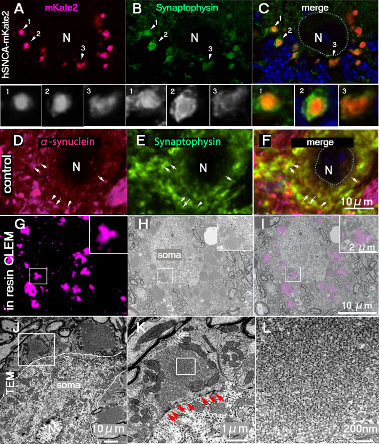
α-Synuclein is the causative gene for PARK1 and PARK4 (heterozygous triplication of SNCA) and is associated with Parkinson's disease, where it localizes to presynaptic terminals in mature neurons. Beyond Parkinson's disease, α-synuclein has also been implicated in various other neuronal disorders. In vitro studies using purified α-synuclein protein have suggested it is involved in synaptic vesicle assembly. However, its physiological function and the ultrastructure of its localization sites in presynaptic terminals remain unclear. To address this, we generated transgenic mice overexpressing human α-synuclein tagged with mKate2 (hSNCA-mKate2 mice) to investigate its in vivo role in synaptic vesicle pool formation at presynaptic terminals. These mice showed normal growth and fertility, and even at 1-yr. old, they showed no motor dysfunction compared to their wild-type littermates. Additionally, no abnormal protein aggregates indicative of neurodegeneration were observed. In this review, we summarize recent findings on the in vivo role of α-synuclein within presynaptic terminals, utilizing hSNCA-mKate2 mice in combination with in-resin correlative light and electron microscopy, electron microscopy, and immunohistochemistry.
期刊介绍:
Acta Histochemica et Cytochemica is the official online journal of the Japan Society of Histochemistry and Cytochemistry. It is intended primarily for rapid publication of concise, original articles in the fields of histochemistry and cytochemistry. Manuscripts oriented towards methodological subjects that contain significant technical advances in these fields are also welcome. Manuscripts in English are accepted from investigators in any country, whether or not they are members of the Japan Society of Histochemistry and Cytochemistry. Manuscripts should be original work that has not been previously published and is not being considered for publication elsewhere, with the exception of abstracts. Manuscripts with essentially the same content as a paper that has been published or accepted, or is under consideration for publication, will not be considered. All submitted papers will be peer-reviewed by at least two referees selected by an appropriate Associate Editor. Acceptance is based on scientific significance, originality, and clarity. When required, a revised manuscript should be submitted within 3 months, otherwise it will be considered to be a new submission. The Editor-in-Chief will make all final decisions regarding acceptance.

 求助内容:
求助内容: 应助结果提醒方式:


