Impact of IKCa Channels on CD34+ Cells in Arteriole Remodeling in Angiotensin II-Induced Hypertension Model Mice.

IF 6.9 1区 医学 Q1 PERIPHERAL VASCULAR DISEASE
Hai Tian,Xin Liao,Cheng Xie,Xiaolin Zhang,Guoqiang Ren,Pengwei Zhu,Yan Yang,Pengyun Li,Changli Liao,Chunshu Li,Qingbo Xu,Xiangyuan Pu,Jun Cheng
{"title":"Impact of IKCa Channels on CD34+ Cells in Arteriole Remodeling in Angiotensin II-Induced Hypertension Model Mice.","authors":"Hai Tian,Xin Liao,Cheng Xie,Xiaolin Zhang,Guoqiang Ren,Pengwei Zhu,Yan Yang,Pengyun Li,Changli Liao,Chunshu Li,Qingbo Xu,Xiangyuan Pu,Jun Cheng","doi":"10.1161/hypertensionaha.124.24537","DOIUrl":null,"url":null,"abstract":"BACKGROUND\r\nMechanisms of endothelial repair in hypertension remain unclear. CD34+ cells are reported to contribute to vascular regeneration; however, their origin and regulation in hypertension are poorly understood. We investigated the role of IKCa channels in CD34+ cell-mediated endothelial repair during Ang II (angiotensin II)-induced arteriole remodeling.\r\n\r\nMETHODS\r\nUsing inducible lineage tracing (Cd34-CreERT2; R26-tdTomato), we tracked nonbone marrow-derived CD34+ cells in hypertensive mice. Single-cell RNA sequencing, immunofluorescence, transwell migration assays, and patch-clamp techniques were used to analyze phenotypic transitions, ion channel activity, and signaling pathways. Bone marrow transplantation, the IKCa channel inhibitor TRAM-34, and the ERK (extracellular signal-regulated kinase) inhibitor PD98059 were used to validate functional mechanisms.\r\n\r\nRESULTS\r\nLineage tracing revealed that nonbone marrow-derived CD34+ cells contributed to endothelial repair under hypertensive conditions. Immunofluorescence analysis showed an increase in CD31+-tdTomato+ cells in the arterioles of Ang II-treated mice after 6 weeks, indicating improved endothelial integrity. Single-cell RNA sequencing revealed 2 subgroups of endothelial cells, one of which expressed stem cell markers such as CD34 (cluster of differentiation 34), Flk-1 (fetal liver kinase 1), and Sca-1 (stem cell antigen-1). Gene expression analysis showed that CD34+ cells are involved in endothelial repair through the regulation of cell migration. Importantly, IKCa channel activation facilitated CD34+ cell migration, and TRAM-34-based inhibition of IKCa channels reduced migration. Mechanistic studies revealed that Ang II enhanced CD34+ cell migration via IKCa-mediated activation of the ERK/P38 signaling pathway, promoting cytoskeletal reorganization and increased intracellular calcium levels.\r\n\r\nCONCLUSIONS\r\nArteriole-resident CD34+ cells contribute to endothelial repair in Ang II-induced hypertension. Moreover, IKCa channel upregulation facilitates CD34+ cell migration via ERK/P38 signaling, suggesting potential therapeutic targets for hypertension.","PeriodicalId":13042,"journal":{"name":"Hypertension","volume":"21 1","pages":""},"PeriodicalIF":6.9000,"publicationDate":"2025-07-09","publicationTypes":"Journal Article","fieldsOfStudy":null,"isOpenAccess":false,"openAccessPdf":"","citationCount":"0","resultStr":null,"platform":"Semanticscholar","paperid":null,"PeriodicalName":"Hypertension","FirstCategoryId":"3","ListUrlMain":"https://doi.org/10.1161/hypertensionaha.124.24537","RegionNum":1,"RegionCategory":"医学","ArticlePicture":[],"TitleCN":null,"AbstractTextCN":null,"PMCID":null,"EPubDate":"","PubModel":"","JCR":"Q1","JCRName":"PERIPHERAL VASCULAR DISEASE","Score":null,"Total":0}
引用次数: 0

Abstract

BACKGROUND Mechanisms of endothelial repair in hypertension remain unclear. CD34+ cells are reported to contribute to vascular regeneration; however, their origin and regulation in hypertension are poorly understood. We investigated the role of IKCa channels in CD34+ cell-mediated endothelial repair during Ang II (angiotensin II)-induced arteriole remodeling. METHODS Using inducible lineage tracing (Cd34-CreERT2; R26-tdTomato), we tracked nonbone marrow-derived CD34+ cells in hypertensive mice. Single-cell RNA sequencing, immunofluorescence, transwell migration assays, and patch-clamp techniques were used to analyze phenotypic transitions, ion channel activity, and signaling pathways. Bone marrow transplantation, the IKCa channel inhibitor TRAM-34, and the ERK (extracellular signal-regulated kinase) inhibitor PD98059 were used to validate functional mechanisms. RESULTS Lineage tracing revealed that nonbone marrow-derived CD34+ cells contributed to endothelial repair under hypertensive conditions. Immunofluorescence analysis showed an increase in CD31+-tdTomato+ cells in the arterioles of Ang II-treated mice after 6 weeks, indicating improved endothelial integrity. Single-cell RNA sequencing revealed 2 subgroups of endothelial cells, one of which expressed stem cell markers such as CD34 (cluster of differentiation 34), Flk-1 (fetal liver kinase 1), and Sca-1 (stem cell antigen-1). Gene expression analysis showed that CD34+ cells are involved in endothelial repair through the regulation of cell migration. Importantly, IKCa channel activation facilitated CD34+ cell migration, and TRAM-34-based inhibition of IKCa channels reduced migration. Mechanistic studies revealed that Ang II enhanced CD34+ cell migration via IKCa-mediated activation of the ERK/P38 signaling pathway, promoting cytoskeletal reorganization and increased intracellular calcium levels. CONCLUSIONS Arteriole-resident CD34+ cells contribute to endothelial repair in Ang II-induced hypertension. Moreover, IKCa channel upregulation facilitates CD34+ cell migration via ERK/P38 signaling, suggesting potential therapeutic targets for hypertension.
IKCa通道对血管紧张素ii诱导的高血压模型小鼠小动脉重构中CD34+细胞的影响
背景:高血压血管内皮修复的机制尚不清楚。据报道,CD34+细胞有助于血管再生;然而,它们在高血压中的起源和调控尚不清楚。我们研究了在Ang II(血管紧张素II)诱导的小动脉重塑过程中,IKCa通道在CD34+细胞介导的内皮修复中的作用。方法采用诱导谱系追踪(Cd34-CreERT2;R26-tdTomato),我们在高血压小鼠中追踪了非骨髓来源的CD34+细胞。单细胞RNA测序、免疫荧光、跨井迁移测定和膜片钳技术用于分析表型转变、离子通道活性和信号通路。通过骨髓移植、IKCa通道抑制剂TRAM-34和ERK(细胞外信号调节激酶)抑制剂PD98059来验证其功能机制。结果谱系追踪显示,非骨髓来源的CD34+细胞有助于高血压病患者的内皮修复。免疫荧光分析显示,6周后,angii处理小鼠的小动脉中CD31+-tdTomato+细胞增加,表明内皮完整性得到改善。单细胞RNA测序显示内皮细胞有2个亚群,其中一个亚群表达干细胞标志物,如CD34(分化簇34)、Flk-1(胎儿肝激酶1)和Sca-1(干细胞抗原-1)。基因表达分析表明,CD34+细胞通过调控细胞迁移参与内皮修复。重要的是,IKCa通道激活促进了CD34+细胞的迁移,而基于tram -34的IKCa通道抑制减少了迁移。机制研究表明,Ang II通过ikca介导的ERK/P38信号通路激活,促进CD34+细胞迁移,促进细胞骨架重组,增加细胞内钙水平。结论sarteriole -驻地CD34+细胞参与了angii诱导的高血压的内皮修复。此外,IKCa通道上调通过ERK/P38信号通路促进CD34+细胞迁移,提示高血压的潜在治疗靶点。
本文章由计算机程序翻译,如有差异,请以英文原文为准。
求助全文
约1分钟内获得全文 求助全文
来源期刊
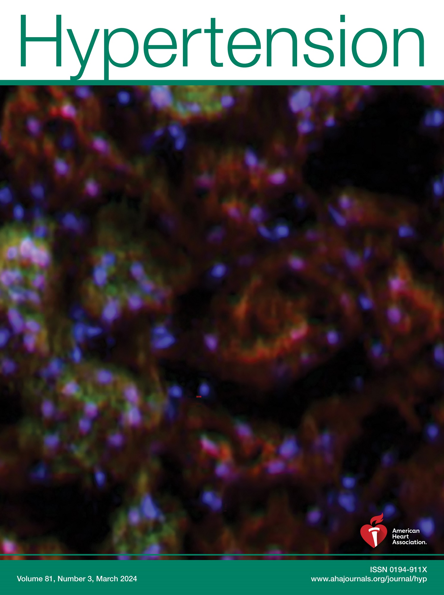
Hypertension
Hypertension 医学-外周血管病
CiteScore
15.90
自引率
4.80%
发文量
1006
审稿时长
1 months
期刊介绍: Hypertension presents top-tier articles on high blood pressure in each monthly release. These articles delve into basic science, clinical treatment, and prevention of hypertension and associated cardiovascular, metabolic, and renal conditions. Renowned for their lasting significance, these papers contribute to advancing our understanding and management of hypertension-related issues.
×
引用
GB/T 7714-2015
复制
MLA
复制
APA
复制
导出至
BibTeX EndNote RefMan NoteFirst NoteExpress
×
提示
您的信息不完整,为了账户安全,请先补充。
现在去补充
×
提示
您因"违规操作"
具体请查看互助需知
我知道了
×
提示
确定
请完成安全验证×
copy
已复制链接
快去分享给好友吧!
我知道了
右上角分享
点击右上角分享
0
联系我们:info@booksci.cn Book学术提供免费学术资源搜索服务,方便国内外学者检索中英文文献。致力于提供最便捷和优质的服务体验。 Copyright © 2023 布克学术 All rights reserved.
京ICP备2023020795号-1
ghs 京公网安备 11010802042870号
Book学术文献互助
Book学术文献互助群
群 号:604180095
Book学术官方微信