Marlena Bereźniak, Krzysztof Piłat, Grzegorz Benke, Jarosław Czerwiński, Marta Byrdy-Daca, Jan Świątkowski, Katarzyna Sułkowska, Paweł Łęgosz, Marek Gołębiowski, Piotr Palczewski
{"title":"Acute thoracolumbar fractures in patients with a rigid spine: a computed tomography study.","authors":"Marlena Bereźniak, Krzysztof Piłat, Grzegorz Benke, Jarosław Czerwiński, Marta Byrdy-Daca, Jan Świątkowski, Katarzyna Sułkowska, Paweł Łęgosz, Marek Gołębiowski, Piotr Palczewski","doi":"10.5114/pjr/203852","DOIUrl":null,"url":null,"abstract":"<p><strong>Purpose: </strong>To assess the influence of long- and short-segment spinal ankylosis on the characteristics and course of acute thoracolumbar fractures.</p><p><strong>Material and methods: </strong>Computed tomography (CT) studies of 372 patients who were diagnosed with acute thoracolumbar spine fracture in our hospital between 2014 and 2020 were retrospectively reviewed. Demographic data, presence or absence of rigid spine conditions, location, and fracture morphology according to the <i>Arbeitsgemeinschaft für Osteosynthesefragen</i> (AO) spine classification were assessed. Statistical analysis was performed using the χ<sup>2</sup> test.</p><p><strong>Results: </strong>A total of 65 patients with fractures through ankylosed segment or immediately adjacent segment (rigid spine group) and 307 controls were identified. Most rigid spine patients suffered minor trauma. In both groups most of the fractures were located in the thoracolumbar junction, and type A1 fractures were most common, followed by types A3 and A4. Multilevel fractures were more common in rigid spine patients (41.54% vs. 30.29%). Most of the rigid spine fractures (46.96%) were located within the fused spinal segment, with the midportion of the fused spinal segment being the most common location of types B and C fractures. Long-segment fusion was associated with unstable type B and C fractures. In short-segment fusion, single level type A fractures were most common. Spinal cord injury occurred only in patients with delayed diagnosis.</p><p><strong>Conclusions: </strong>When plain films are used as a first-line diagnostic test for thoracolumbar spine trauma in stable patients without abnormal neurological signs or symptoms, and long-segment spinal ankylosis is observed, thoracolumbar CT should be used for further evaluation.</p>","PeriodicalId":94174,"journal":{"name":"Polish journal of radiology","volume":"90 ","pages":"e215-e223"},"PeriodicalIF":0.0000,"publicationDate":"2025-05-09","publicationTypes":"Journal Article","fieldsOfStudy":null,"isOpenAccess":false,"openAccessPdf":"https://www.ncbi.nlm.nih.gov/pmc/articles/PMC12232406/pdf/","citationCount":"0","resultStr":null,"platform":"Semanticscholar","paperid":null,"PeriodicalName":"Polish journal of radiology","FirstCategoryId":"1085","ListUrlMain":"https://doi.org/10.5114/pjr/203852","RegionNum":0,"RegionCategory":null,"ArticlePicture":[],"TitleCN":null,"AbstractTextCN":null,"PMCID":null,"EPubDate":"2025/1/1 0:00:00","PubModel":"eCollection","JCR":"","JCRName":"","Score":null,"Total":0}
引用次数: 0
Abstract
Purpose: To assess the influence of long- and short-segment spinal ankylosis on the characteristics and course of acute thoracolumbar fractures.
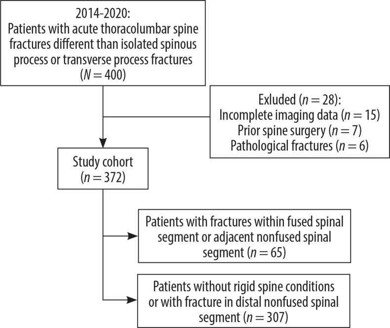
Material and methods: Computed tomography (CT) studies of 372 patients who were diagnosed with acute thoracolumbar spine fracture in our hospital between 2014 and 2020 were retrospectively reviewed. Demographic data, presence or absence of rigid spine conditions, location, and fracture morphology according to the Arbeitsgemeinschaft für Osteosynthesefragen (AO) spine classification were assessed. Statistical analysis was performed using the χ2 test.
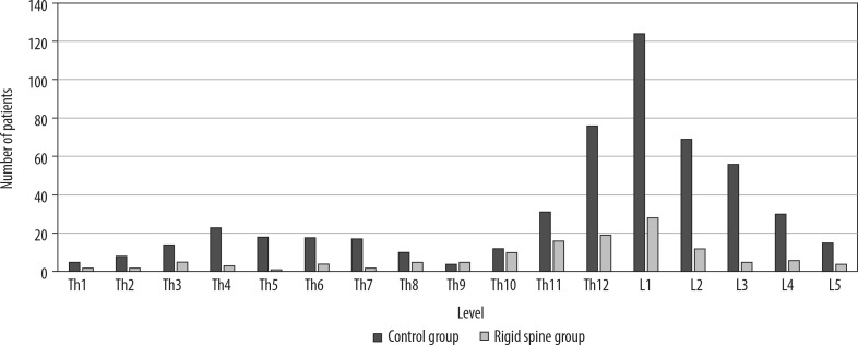
Results: A total of 65 patients with fractures through ankylosed segment or immediately adjacent segment (rigid spine group) and 307 controls were identified. Most rigid spine patients suffered minor trauma. In both groups most of the fractures were located in the thoracolumbar junction, and type A1 fractures were most common, followed by types A3 and A4. Multilevel fractures were more common in rigid spine patients (41.54% vs. 30.29%). Most of the rigid spine fractures (46.96%) were located within the fused spinal segment, with the midportion of the fused spinal segment being the most common location of types B and C fractures. Long-segment fusion was associated with unstable type B and C fractures. In short-segment fusion, single level type A fractures were most common. Spinal cord injury occurred only in patients with delayed diagnosis.
Conclusions: When plain films are used as a first-line diagnostic test for thoracolumbar spine trauma in stable patients without abnormal neurological signs or symptoms, and long-segment spinal ankylosis is observed, thoracolumbar CT should be used for further evaluation.

 求助内容:
求助内容: 应助结果提醒方式:


