Assessment of upper airway and temporomandibular joint changes in growing patients with Class II Division 1 malocclusion, treated with the Twin Block appliance: a retrospective cone-beam computed tomography study.
Ioannis Anagnostopoulos, Snigdha Pattanaik, Ahmed Zaky, Ahmed El-Motayam, Shishir Ram Shetty, Mais M Sadek
{"title":"Assessment of upper airway and temporomandibular joint changes in growing patients with Class II Division 1 malocclusion, treated with the Twin Block appliance: a retrospective cone-beam computed tomography study.","authors":"Ioannis Anagnostopoulos, Snigdha Pattanaik, Ahmed Zaky, Ahmed El-Motayam, Shishir Ram Shetty, Mais M Sadek","doi":"10.7181/acfs.2024.0093","DOIUrl":null,"url":null,"abstract":"<p><strong>Background: </strong>The Twin Block (TWB) appliance is widely employed for treating Class II malocclusion in children and adolescents. This study aimed to evaluate the three-dimensional treatment effects of the TWB on the upper airway and temporomandibular joint (TMJ), and to investigate the association between airway changes and TMJ alterations.</p><p><strong>Methods: </strong>This retrospective study examined 24 cone-beam computed tomography (CBCT) scans from 12 patients (mean age, 12.30 ± 1.24 years) diagnosed with Class II Division 1 malocclusion and treated with the TWB appliance. CBCT scans were acquired both at pretreatment (T0) and posttreatment (T1). Romexis 6.2.1 imaging software was used to assess changes in the upper airway and TMJ. The paired t-test was used to compare the pretreatment and posttreatment measurements, while Pearson correlation coefficient analysis evaluated the relationship between the upper airway and TMJ measurements.</p><p><strong>Results: </strong>A statistically significant increase was observed in the upper airway volume, condylar volume, and condylar dimensions after treatment. No significant correlation was detected between the upper airway and TMJ measurements at T0, T1, or in the net changes (T1-T0) during TWB therapy.</p><p><strong>Conclusion: </strong>Growing patients treated with the TWB appliance demonstrated a statistically significant increase in upper airway volume. In addition, there was a significant increase in condylar volume, width, and length, with the condyle repositioned more anteriorly within the glenoid fossa. However, no statistically significant correlation was found between the TMJ and upper airway measurements.</p>","PeriodicalId":52238,"journal":{"name":"Archives of Craniofacial Surgery","volume":"26 3","pages":"92-101"},"PeriodicalIF":0.0000,"publicationDate":"2025-06-01","publicationTypes":"Journal Article","fieldsOfStudy":null,"isOpenAccess":false,"openAccessPdf":"https://www.ncbi.nlm.nih.gov/pmc/articles/PMC12235306/pdf/","citationCount":"0","resultStr":null,"platform":"Semanticscholar","paperid":null,"PeriodicalName":"Archives of Craniofacial Surgery","FirstCategoryId":"1085","ListUrlMain":"https://doi.org/10.7181/acfs.2024.0093","RegionNum":0,"RegionCategory":null,"ArticlePicture":[],"TitleCN":null,"AbstractTextCN":null,"PMCID":null,"EPubDate":"2025/6/20 0:00:00","PubModel":"Epub","JCR":"Q2","JCRName":"Medicine","Score":null,"Total":0}
引用次数: 0
Abstract
Background: The Twin Block (TWB) appliance is widely employed for treating Class II malocclusion in children and adolescents. This study aimed to evaluate the three-dimensional treatment effects of the TWB on the upper airway and temporomandibular joint (TMJ), and to investigate the association between airway changes and TMJ alterations.
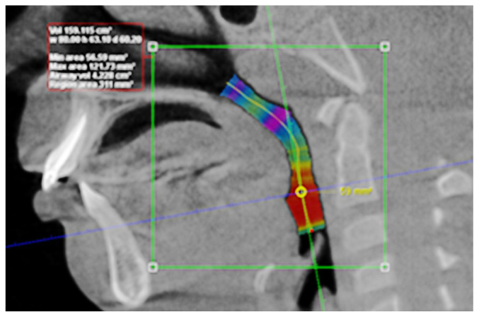
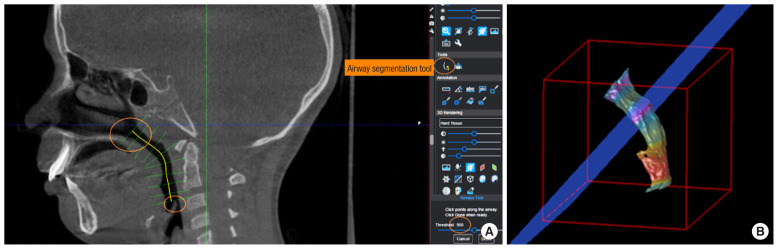
Methods: This retrospective study examined 24 cone-beam computed tomography (CBCT) scans from 12 patients (mean age, 12.30 ± 1.24 years) diagnosed with Class II Division 1 malocclusion and treated with the TWB appliance. CBCT scans were acquired both at pretreatment (T0) and posttreatment (T1). Romexis 6.2.1 imaging software was used to assess changes in the upper airway and TMJ. The paired t-test was used to compare the pretreatment and posttreatment measurements, while Pearson correlation coefficient analysis evaluated the relationship between the upper airway and TMJ measurements.
Results: A statistically significant increase was observed in the upper airway volume, condylar volume, and condylar dimensions after treatment. No significant correlation was detected between the upper airway and TMJ measurements at T0, T1, or in the net changes (T1-T0) during TWB therapy.
Conclusion: Growing patients treated with the TWB appliance demonstrated a statistically significant increase in upper airway volume. In addition, there was a significant increase in condylar volume, width, and length, with the condyle repositioned more anteriorly within the glenoid fossa. However, no statistically significant correlation was found between the TMJ and upper airway measurements.

 求助内容:
求助内容: 应助结果提醒方式:


