Farbod Khorrami, Neeru Gupta, Xun Zhou, You Liang, Yeni H Yucel
{"title":"A Novel Retinal Nerve Fiber Layer Biomarker of Amyotrophic Lateral Sclerosis (ALS) Identified Using Longitudinal in vivo Ocular Imaging.","authors":"Farbod Khorrami, Neeru Gupta, Xun Zhou, You Liang, Yeni H Yucel","doi":"10.2147/EB.S516163","DOIUrl":null,"url":null,"abstract":"<p><strong>Purpose: </strong>Like motor neurons, retinal ganglion cells (RGCs) have long axons and high metabolic demands, making them vulnerable to disruption of axonal transport. Unlike motor neurons, the RGC axons are accessible to high-resolution non-invasive optical imaging in their intraocular portion. A non-invasive in vivo retinal imaging biomarker can be valuable for amyotrophic lateral sclerosis (ALS) diagnosis and monitoring. We aim to assess the presence of inner retinal pathology in a mouse model of ALS and its possible progression with age.</p><p><strong>Methods: </strong>Transgenic SOD1G93A mice (n=8, 4M/4F) and age-matched controls (n=8, 4M/4F) underwent in vivo retinal imaging with confocal scanning laser ophthalmoscopy (cSLO) coupled with optical coherence tomography (OCT) at 20 weeks of age. Another group of SOD1G93A mice (n=20, 6M/14F) and age-matched controls (n=20, 6M/14F) underwent longitudinal in vivo retinal imaging with the same device. Each retinal imaging session included infrared reflectance (IR) and blue reflectance (BR) cSLO coupled with OCT. Hyperreflective puncta located in the retinal nerve fiber layer (RNFL) were counted in a blinded fashion in ALS and control mice. The number of puncta at 20 weeks of age in ALS mice was compared with controls using Wilcoxon test. The rates of increase of puncta number were analyzed using a Generalized Linear Mixed-Effect Model (GLMM) for genotype, time, and sex.</p><p><strong>Results: </strong>IR-cSLO coupled with OCT revealed hyperreflective puncta located in the RNFL of ALS mice. IR-cSLO fundus imaging at the age of 20 weeks showed ALS mice had significantly higher number of puncta compared to controls (2.1±2.3 vs 0.5±0.8; (mean±SD), respectively, p=0.036). GLMM analysis showed both ALS mutation and age were significantly associated with the rate of increase of puncta number (p=0.000232 and p=0.000366, respectively). In addition, female ALS mice had a steeper increase of puncta compared to male ALS mice (0.21±0.04 log number puncta/week vs 0.16±0.04, respectively; p=0.037).</p><p><strong>Conclusion: </strong>Our findings demonstrate distinct inner retinal nerve fiber layer pathology, detected using cSLO coupled with OCT, which worsens over time. These findings support the potential of retinal imaging as a translationally relevant, non-invasive biomarker for ALS diagnosis or disease monitoring in humans.</p>","PeriodicalId":51844,"journal":{"name":"Eye and Brain","volume":"17 ","pages":"69-79"},"PeriodicalIF":2.4000,"publicationDate":"2025-07-02","publicationTypes":"Journal Article","fieldsOfStudy":null,"isOpenAccess":false,"openAccessPdf":"https://www.ncbi.nlm.nih.gov/pmc/articles/PMC12230754/pdf/","citationCount":"0","resultStr":null,"platform":"Semanticscholar","paperid":null,"PeriodicalName":"Eye and Brain","FirstCategoryId":"1085","ListUrlMain":"https://doi.org/10.2147/EB.S516163","RegionNum":0,"RegionCategory":null,"ArticlePicture":[],"TitleCN":null,"AbstractTextCN":null,"PMCID":null,"EPubDate":"2025/1/1 0:00:00","PubModel":"eCollection","JCR":"Q1","JCRName":"OPHTHALMOLOGY","Score":null,"Total":0}
引用次数: 0
Abstract
Purpose: Like motor neurons, retinal ganglion cells (RGCs) have long axons and high metabolic demands, making them vulnerable to disruption of axonal transport. Unlike motor neurons, the RGC axons are accessible to high-resolution non-invasive optical imaging in their intraocular portion. A non-invasive in vivo retinal imaging biomarker can be valuable for amyotrophic lateral sclerosis (ALS) diagnosis and monitoring. We aim to assess the presence of inner retinal pathology in a mouse model of ALS and its possible progression with age.
Methods: Transgenic SOD1G93A mice (n=8, 4M/4F) and age-matched controls (n=8, 4M/4F) underwent in vivo retinal imaging with confocal scanning laser ophthalmoscopy (cSLO) coupled with optical coherence tomography (OCT) at 20 weeks of age. Another group of SOD1G93A mice (n=20, 6M/14F) and age-matched controls (n=20, 6M/14F) underwent longitudinal in vivo retinal imaging with the same device. Each retinal imaging session included infrared reflectance (IR) and blue reflectance (BR) cSLO coupled with OCT. Hyperreflective puncta located in the retinal nerve fiber layer (RNFL) were counted in a blinded fashion in ALS and control mice. The number of puncta at 20 weeks of age in ALS mice was compared with controls using Wilcoxon test. The rates of increase of puncta number were analyzed using a Generalized Linear Mixed-Effect Model (GLMM) for genotype, time, and sex.
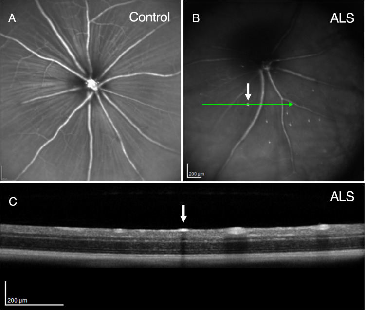
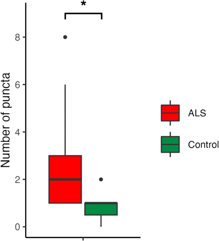
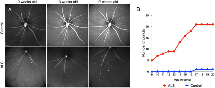
Results: IR-cSLO coupled with OCT revealed hyperreflective puncta located in the RNFL of ALS mice. IR-cSLO fundus imaging at the age of 20 weeks showed ALS mice had significantly higher number of puncta compared to controls (2.1±2.3 vs 0.5±0.8; (mean±SD), respectively, p=0.036). GLMM analysis showed both ALS mutation and age were significantly associated with the rate of increase of puncta number (p=0.000232 and p=0.000366, respectively). In addition, female ALS mice had a steeper increase of puncta compared to male ALS mice (0.21±0.04 log number puncta/week vs 0.16±0.04, respectively; p=0.037).
Conclusion: Our findings demonstrate distinct inner retinal nerve fiber layer pathology, detected using cSLO coupled with OCT, which worsens over time. These findings support the potential of retinal imaging as a translationally relevant, non-invasive biomarker for ALS diagnosis or disease monitoring in humans.
期刊介绍:
Eye and Brain is an international, peer-reviewed, open access journal focusing on basic research, clinical findings, and expert reviews in the field of visual science and neuro-ophthalmology. The journal’s unique focus is the link between two well-known visual centres, the eye and the brain, with an emphasis on the importance of such connections. All aspects of clinical and especially basic research on the visual system are addressed within the journal as well as significant future directions in vision research and therapeutic measures. This unique journal focuses on neurological aspects of vision – both physiological and pathological. The scope of the journal spans from the cornea to the associational visual cortex and all the visual centers in between. Topics range from basic biological mechanisms to therapeutic treatment, from simple organisms to humans, and utilizing techniques from molecular biology to behavior. The journal especially welcomes primary research articles or review papers that make the connection between the eye and the brain. Specific areas covered in the journal include: Physiology and pathophysiology of visual centers, Eye movement disorders and strabismus, Cellular, biochemical, and molecular features of the visual system, Structural and functional organization of the eye and of the visual cortex, Metabolic demands of the visual system, Diseases and disorders with neuro-ophthalmic manifestations, Clinical and experimental neuro-ophthalmology and visual system pathologies, Epidemiological studies.

 求助内容:
求助内容: 应助结果提醒方式:


