{"title":"Construction of a prediction model for peripheral lymph node metastasis in patients with colorectal cancer based on enhanced CT texture features.","authors":"Feng Tong, Longfei Zhang, Xiaobin Jiang, Zhenyu Wu","doi":"10.1186/s12957-025-03928-6","DOIUrl":null,"url":null,"abstract":"<p><strong>Background: </strong>To investigate the analysis of peripheral lymph node metastasis prediction model construction for patients with colorectal cancer based on enhanced CT texture features.</p><p><strong>Methods: </strong>In this study, the clinical data of 200 colorectal cancer patients admitted to our hospital from January 2019 to October 2024 were collected, which were divided into a training set (n = 140) and a validation set (n = 60) according to a 7:3 ratio. The training set was used to construct the prediction model and the validation set was used to evaluate the model performance. Independent influencing factors of peripheral lymph node metastasis in colorectal cancer patients were screened by single-factor and multifactor logistic regression analyses, and the prediction model was finally constructed and analysed for its predictive effect using ROC curves and decision curves.</p><p><strong>Results: </strong>In the training and validation sets, compared with those without lymph node metastasis, colorectal cancer patients with lymph node metastasis had a higher percentage of those whose tumour infiltration depth was submucosal and those whose tumour differentiation was poorly differentiated, and the skewness, kurtosis, and entropy values of their CT texture features were also significantly higher than those without lymph node metastasis (P < 0.05). Multifactorial logistic regression analysis showed that the depth of tumour infiltration was higher for submucosal layer (OR = 3.367, 95% CI = 1.104 ~ 1.271), tumour hypofractionation (OR = 3.881, 95% CI = 1.04714.392), skewness (OR = 3.979, 95% CI = 1.04714.392), kurtosis (OR = 4.824, 95% CI = 2.251 ~ 10.336), and entropy (OR = 2.221, 95% CI = 1.159 ~ 4.257) were independent risk factors for lymph node metastasis in colorectal cancer patients. The consistency index (C-index) of the lymph node metastasis prediction model based on enhanced CT texture features was 0.980, and the calibration curve results were basically consistent with the predicted values; the AUCs of lymph node metastasis prediction for the training and validation sets were 0.937 and 0.960, respectively. Decision curve analysis showed that the clinical decision-making benefit of the model was significantly improved after adding CT texture features.</p><p><strong>Conclusion: </strong>The prediction model based on enhanced CT texture features has good predictive value for predicting peripheral lymph node metastasis in colorectal cancer.</p>","PeriodicalId":23856,"journal":{"name":"World Journal of Surgical Oncology","volume":"23 1","pages":"266"},"PeriodicalIF":2.5000,"publicationDate":"2025-07-07","publicationTypes":"Journal Article","fieldsOfStudy":null,"isOpenAccess":false,"openAccessPdf":"https://www.ncbi.nlm.nih.gov/pmc/articles/PMC12236036/pdf/","citationCount":"0","resultStr":null,"platform":"Semanticscholar","paperid":null,"PeriodicalName":"World Journal of Surgical Oncology","FirstCategoryId":"3","ListUrlMain":"https://doi.org/10.1186/s12957-025-03928-6","RegionNum":3,"RegionCategory":"医学","ArticlePicture":[],"TitleCN":null,"AbstractTextCN":null,"PMCID":null,"EPubDate":"","PubModel":"","JCR":"Q3","JCRName":"ONCOLOGY","Score":null,"Total":0}
引用次数: 0
Abstract
Background: To investigate the analysis of peripheral lymph node metastasis prediction model construction for patients with colorectal cancer based on enhanced CT texture features.
Methods: In this study, the clinical data of 200 colorectal cancer patients admitted to our hospital from January 2019 to October 2024 were collected, which were divided into a training set (n = 140) and a validation set (n = 60) according to a 7:3 ratio. The training set was used to construct the prediction model and the validation set was used to evaluate the model performance. Independent influencing factors of peripheral lymph node metastasis in colorectal cancer patients were screened by single-factor and multifactor logistic regression analyses, and the prediction model was finally constructed and analysed for its predictive effect using ROC curves and decision curves.
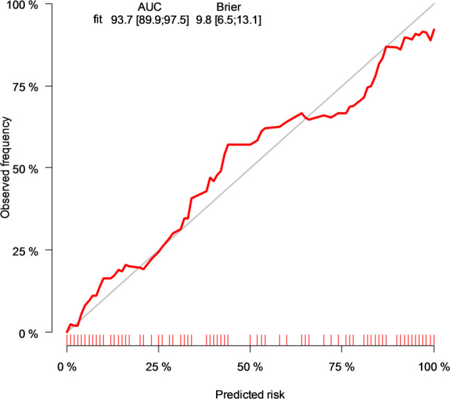
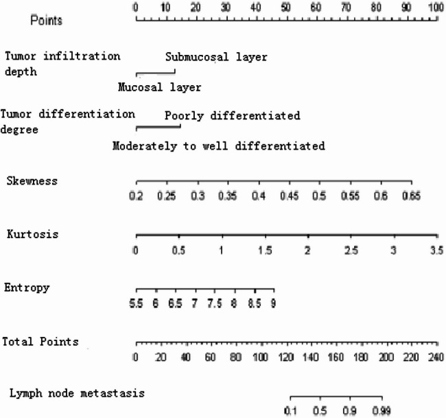
Results: In the training and validation sets, compared with those without lymph node metastasis, colorectal cancer patients with lymph node metastasis had a higher percentage of those whose tumour infiltration depth was submucosal and those whose tumour differentiation was poorly differentiated, and the skewness, kurtosis, and entropy values of their CT texture features were also significantly higher than those without lymph node metastasis (P < 0.05). Multifactorial logistic regression analysis showed that the depth of tumour infiltration was higher for submucosal layer (OR = 3.367, 95% CI = 1.104 ~ 1.271), tumour hypofractionation (OR = 3.881, 95% CI = 1.04714.392), skewness (OR = 3.979, 95% CI = 1.04714.392), kurtosis (OR = 4.824, 95% CI = 2.251 ~ 10.336), and entropy (OR = 2.221, 95% CI = 1.159 ~ 4.257) were independent risk factors for lymph node metastasis in colorectal cancer patients. The consistency index (C-index) of the lymph node metastasis prediction model based on enhanced CT texture features was 0.980, and the calibration curve results were basically consistent with the predicted values; the AUCs of lymph node metastasis prediction for the training and validation sets were 0.937 and 0.960, respectively. Decision curve analysis showed that the clinical decision-making benefit of the model was significantly improved after adding CT texture features.
Conclusion: The prediction model based on enhanced CT texture features has good predictive value for predicting peripheral lymph node metastasis in colorectal cancer.
期刊介绍:
World Journal of Surgical Oncology publishes articles related to surgical oncology and its allied subjects, such as epidemiology, cancer research, biomarkers, prevention, pathology, radiology, cancer treatment, clinical trials, multimodality treatment and molecular biology. Emphasis is placed on original research articles. The journal also publishes significant clinical case reports, as well as balanced and timely reviews on selected topics.
Oncology is a multidisciplinary super-speciality of which surgical oncology forms an integral component, especially with solid tumors. Surgical oncologists around the world are involved in research extending from detecting the mechanisms underlying the causation of cancer, to its treatment and prevention. The role of a surgical oncologist extends across the whole continuum of care. With continued developments in diagnosis and treatment, the role of a surgical oncologist is ever-changing. Hence, World Journal of Surgical Oncology aims to keep readers abreast with latest developments that will ultimately influence the work of surgical oncologists.

 求助内容:
求助内容: 应助结果提醒方式:


