Human papillomavirus (HPV) genotyping and prognostic value of HPV E4 protein and transcription factors NANOG and SOX11 in atypical p16 patchy squamous epithelium of cervix.
Maja Kebe Radulovic, Anja Ostrbenk, Mario Poljak, Margareta Strojan-Flezar
{"title":"Human papillomavirus (HPV) genotyping and prognostic value of HPV E4 protein and transcription factors NANOG and SOX11 in atypical p16 patchy squamous epithelium of cervix.","authors":"Maja Kebe Radulovic, Anja Ostrbenk, Mario Poljak, Margareta Strojan-Flezar","doi":"10.2478/raon-2025-0038","DOIUrl":null,"url":null,"abstract":"<p><strong>Background: </strong>Immunohistochemical staining for p16 is used to differentiate precancerous cervical lesions in tissue samples, but the interpretation of patchy p16 expression remains challenging. We performed human papillomavirus (HPV) genotyping and evaluated immunohistochemical expression of HPV E4 protein - a marker for transient infections, stem cell transcription factor NANOG, and transcription factor SOX11 to detect possible high-grade squamous lesions in atypical p16 patchy squamous epithelium.</p><p><strong>Materials and methods: </strong>We analyzed 24 cervical tissue samples with atypical squamous epithelium and patchy p16 expression along with the following controls: 11 cases of atypical squamous epithelium with null p16 expression, 9 condylomas, 12 cases of cervical intraepithelial neoplasia (CIN) grade 1, 11 cases of CIN2, and 9 cases of CIN3. In addition, HPV genotyping of tissue and related cervical smears from up to two years prior to biopsy was performed. Immunohistochemical staining for Ki67, HPV E4, NANOG, and SOX11 was performed and compared with follow-up data.</p><p><strong>Results: </strong>High-risk HPV infection was detected in 6/24 cases with patchy p16 expression and HPV E4 was expressed in 1/24 cases with patchy p16, weak NANOG expression was found in 11/24 cases with patchy p16 expression while no SOX11 expression was observed. During 10 months of follow-up, additional CIN1 and two CIN3 were identified, and another CIN1 and CIN3 after 5 and 6 years, accordingly.</p><p><strong>Conclusions: </strong>Our study showed that atypical squamous epithelium with patchy p16 expression poses a risk for highgrade precancerous lesions, harbouring high-risk HPV infection. Novel markers may hold diagnostic value in other specific contexts.</p>","PeriodicalId":21034,"journal":{"name":"Radiology and Oncology","volume":" ","pages":"391-402"},"PeriodicalIF":2.2000,"publicationDate":"2025-07-08","publicationTypes":"Journal Article","fieldsOfStudy":null,"isOpenAccess":false,"openAccessPdf":"https://www.ncbi.nlm.nih.gov/pmc/articles/PMC12441885/pdf/","citationCount":"0","resultStr":null,"platform":"Semanticscholar","paperid":null,"PeriodicalName":"Radiology and Oncology","FirstCategoryId":"3","ListUrlMain":"https://doi.org/10.2478/raon-2025-0038","RegionNum":4,"RegionCategory":"医学","ArticlePicture":[],"TitleCN":null,"AbstractTextCN":null,"PMCID":null,"EPubDate":"2025/9/1 0:00:00","PubModel":"eCollection","JCR":"Q3","JCRName":"ONCOLOGY","Score":null,"Total":0}
引用次数: 0
Abstract
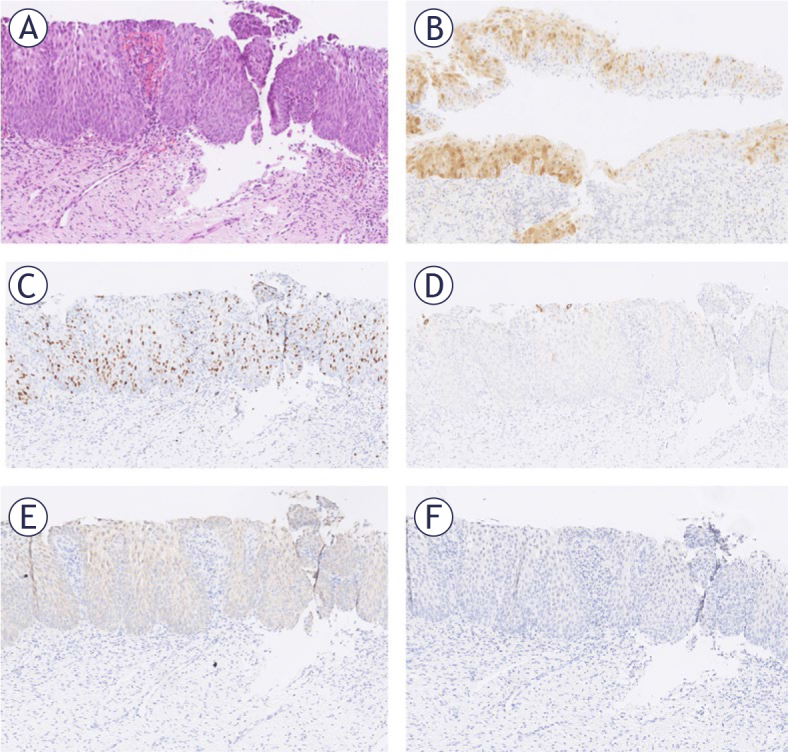
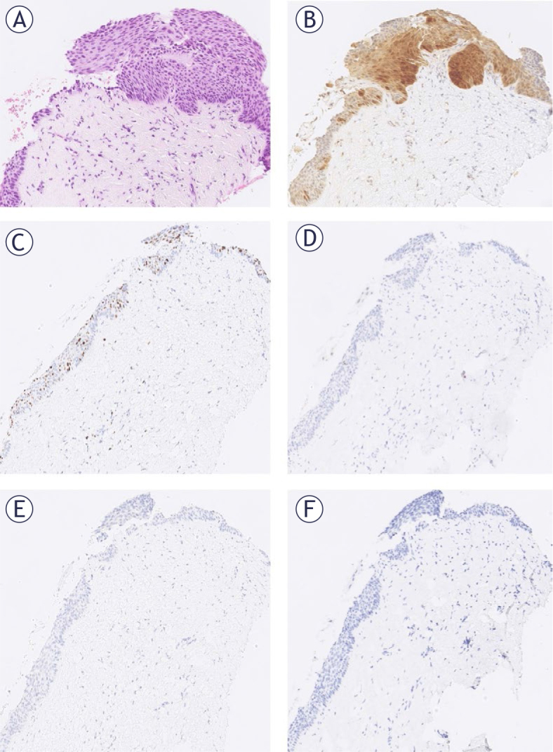
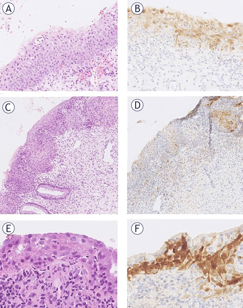
Background: Immunohistochemical staining for p16 is used to differentiate precancerous cervical lesions in tissue samples, but the interpretation of patchy p16 expression remains challenging. We performed human papillomavirus (HPV) genotyping and evaluated immunohistochemical expression of HPV E4 protein - a marker for transient infections, stem cell transcription factor NANOG, and transcription factor SOX11 to detect possible high-grade squamous lesions in atypical p16 patchy squamous epithelium.
Materials and methods: We analyzed 24 cervical tissue samples with atypical squamous epithelium and patchy p16 expression along with the following controls: 11 cases of atypical squamous epithelium with null p16 expression, 9 condylomas, 12 cases of cervical intraepithelial neoplasia (CIN) grade 1, 11 cases of CIN2, and 9 cases of CIN3. In addition, HPV genotyping of tissue and related cervical smears from up to two years prior to biopsy was performed. Immunohistochemical staining for Ki67, HPV E4, NANOG, and SOX11 was performed and compared with follow-up data.
Results: High-risk HPV infection was detected in 6/24 cases with patchy p16 expression and HPV E4 was expressed in 1/24 cases with patchy p16, weak NANOG expression was found in 11/24 cases with patchy p16 expression while no SOX11 expression was observed. During 10 months of follow-up, additional CIN1 and two CIN3 were identified, and another CIN1 and CIN3 after 5 and 6 years, accordingly.
Conclusions: Our study showed that atypical squamous epithelium with patchy p16 expression poses a risk for highgrade precancerous lesions, harbouring high-risk HPV infection. Novel markers may hold diagnostic value in other specific contexts.
期刊介绍:
Radiology and Oncology is a multidisciplinary journal devoted to the publishing original and high quality scientific papers and review articles, pertinent to diagnostic and interventional radiology, computerized tomography, magnetic resonance, ultrasound, nuclear medicine, radiotherapy, clinical and experimental oncology, radiobiology, medical physics and radiation protection. Therefore, the scope of the journal is to cover beside radiology the diagnostic and therapeutic aspects in oncology, which distinguishes it from other journals in the field.
 求助内容:
求助内容: 应助结果提醒方式:


