Comparative Effects of Maxillary Advancement Alone and in Combination with Mandibular Setback on Airway Anatomy and Function in Class III Malocclusion: A Controlled Prospective Clinical Study.
{"title":"Comparative Effects of Maxillary Advancement Alone and in Combination with Mandibular Setback on Airway Anatomy and Function in Class III Malocclusion: A Controlled Prospective Clinical Study.","authors":"Hatice Başaran Bal, Celal Irgın","doi":"10.4274/TurkJOrthod.2025.2025.28","DOIUrl":null,"url":null,"abstract":"<p><strong>Objective: </strong>The aim of this study is to evaluate the effects of maxillary advancement (MxA) and bimaxillary osteotomy (MdS-MxA) on upper pharyngeal airway volume (PAV), apnea-hypopnea index (AHI), hyoid bone (HB) position, and head posture (HP) in young and healthy individuals with skeletal Class III malocclusion.</p><p><strong>Methods: </strong>This prospective clinical study included three groups: MxA, MdS-MxA, and Class I control group, with 12 subjects each. In the surgical groups, lateral cephalometric radiographs, cone-beam computed tomography images, and AHI measurements were obtained preoperatively and approximately six months postoperatively. Only pre-treatment records were collected for the control group. Depending on data distribution, parametric (Paired Samples t-test and ANOVA) or non-parametric (Wilcoxon Signed-Rank and Kruskal-Wallis) tests were used for intra- and inter-group statistical comparisons, with a significance level set at p<0.05.</p><p><strong>Results: </strong>The maxillary forward movement for the MxA group was 5.34 mm. It was 5.32 mm in the MdS-MxA group, and the mandibular setback was 4.71 mm. Nearly six months after surgery, significant differences were observed among the groups in the sagittal positions of the jaws, the vertical position of the mandible, the vertical position of the hyoid bone, and PAV sections. No significant differences were found in HP, minimum cross-sectional area or AHI.</p><p><strong>Conclusion: </strong>PAV increase was observed in both surgical groups. MdS-MxA did not have an effect on obstructive sleep apnea. Postoperative HB displacement was minimal, with a slight inferior shift observed in the MdS-MxA group.</p>","PeriodicalId":37013,"journal":{"name":"Turkish Journal of Orthodontics","volume":"38 2","pages":"107-115"},"PeriodicalIF":1.4000,"publicationDate":"2025-07-02","publicationTypes":"Journal Article","fieldsOfStudy":null,"isOpenAccess":false,"openAccessPdf":"https://www.ncbi.nlm.nih.gov/pmc/articles/PMC12236119/pdf/","citationCount":"0","resultStr":null,"platform":"Semanticscholar","paperid":null,"PeriodicalName":"Turkish Journal of Orthodontics","FirstCategoryId":"1085","ListUrlMain":"https://doi.org/10.4274/TurkJOrthod.2025.2025.28","RegionNum":0,"RegionCategory":null,"ArticlePicture":[],"TitleCN":null,"AbstractTextCN":null,"PMCID":null,"EPubDate":"","PubModel":"","JCR":"Q4","JCRName":"DENTISTRY, ORAL SURGERY & MEDICINE","Score":null,"Total":0}
引用次数: 0
Abstract
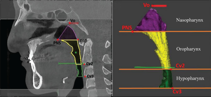
Objective: The aim of this study is to evaluate the effects of maxillary advancement (MxA) and bimaxillary osteotomy (MdS-MxA) on upper pharyngeal airway volume (PAV), apnea-hypopnea index (AHI), hyoid bone (HB) position, and head posture (HP) in young and healthy individuals with skeletal Class III malocclusion.
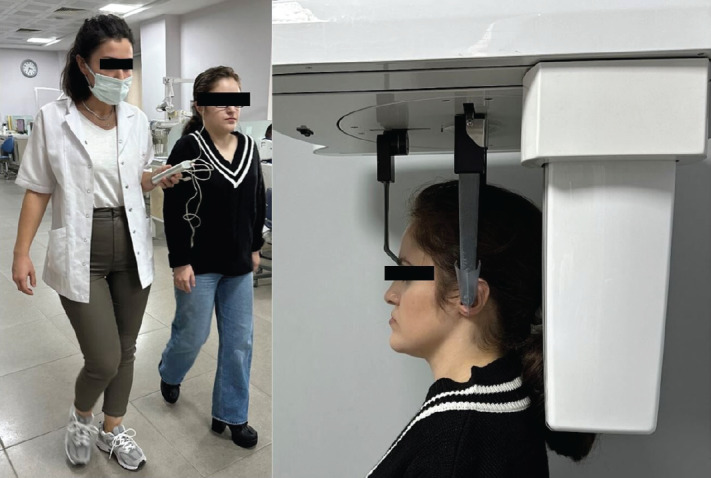
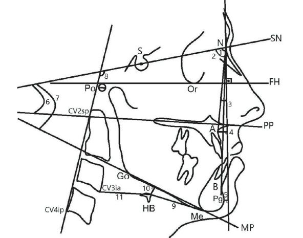
Methods: This prospective clinical study included three groups: MxA, MdS-MxA, and Class I control group, with 12 subjects each. In the surgical groups, lateral cephalometric radiographs, cone-beam computed tomography images, and AHI measurements were obtained preoperatively and approximately six months postoperatively. Only pre-treatment records were collected for the control group. Depending on data distribution, parametric (Paired Samples t-test and ANOVA) or non-parametric (Wilcoxon Signed-Rank and Kruskal-Wallis) tests were used for intra- and inter-group statistical comparisons, with a significance level set at p<0.05.
Results: The maxillary forward movement for the MxA group was 5.34 mm. It was 5.32 mm in the MdS-MxA group, and the mandibular setback was 4.71 mm. Nearly six months after surgery, significant differences were observed among the groups in the sagittal positions of the jaws, the vertical position of the mandible, the vertical position of the hyoid bone, and PAV sections. No significant differences were found in HP, minimum cross-sectional area or AHI.
Conclusion: PAV increase was observed in both surgical groups. MdS-MxA did not have an effect on obstructive sleep apnea. Postoperative HB displacement was minimal, with a slight inferior shift observed in the MdS-MxA group.

 求助内容:
求助内容: 应助结果提醒方式:


