Gilberto Siebert Filho, Helder Fernandes de Oliveira, Alline Soares Vaz, Karolina Kellen Matias, Orlando Aguirre Guedes, Jesus Djalma Pécora, Álvaro Henrique Borges, Rafael Ratto de Moraes
{"title":"Residual dentin thickness in maxillary first premolars with palatal groove after endodontic and restorative procedures: An e-Vol DX analysis.","authors":"Gilberto Siebert Filho, Helder Fernandes de Oliveira, Alline Soares Vaz, Karolina Kellen Matias, Orlando Aguirre Guedes, Jesus Djalma Pécora, Álvaro Henrique Borges, Rafael Ratto de Moraes","doi":"10.4317/jced.62794","DOIUrl":null,"url":null,"abstract":"<p><strong>Background: </strong>This study aimed to measure residual dentin thickness in maxillary first premolars with a palatal groove after root canal instrumentation, filling material removal, and post space preparation using e-Vol DX, an advanced CBCT imaging analysis software.</p><p><strong>Material and methods: </strong>Fourteen extracted maxillary first premolars with a palatal groove on the buccal root were selected. Dentin thickness was measured at 4 stages: initial (M1), after instrumentation (M2), after filling material removal (M3), and after post space preparation (M4). Measurements were taken in 3 root regions: cervical (1 mm coronal to the groove), middle (at the groove's deepest point), and apical (2 mm apical to the groove). Data were analyzed using ANOVA and Tukey's post hoc test (α = 5%).</p><p><strong>Results: </strong>Significant reductions in dentin thickness were found in all walls across treatment phases. In the palatal wall, dentin thickness dropped below 1 mm after instrumentation, retreatment, and post space preparation. In the buccal and mesial walls, thickness fell below 1 mm after retreatment and/or post space preparation. The distal wall was less affected. The average groove depth was 0.66 ± 0.20 mm, and average groove length was 5.72 ± 1.65 mm.</p><p><strong>Conclusions: </strong>Post space preparation in maxillary first premolars with palatal groove on the buccal root significantly reduces dentin thickness, especially in the palatal wall, increasing the risk of root weakening. Clinicians should carefully assess the indication of intraradicular posts in such cases to avoid potential complications. <b>Key words:</b>Cone-beam computed tomography, e-vol DX software, Maxillary first premolar, residual dentin thickness, root thickness.</p>","PeriodicalId":15376,"journal":{"name":"Journal of Clinical and Experimental Dentistry","volume":"17 6","pages":"e705-e712"},"PeriodicalIF":0.0000,"publicationDate":"2025-06-01","publicationTypes":"Journal Article","fieldsOfStudy":null,"isOpenAccess":false,"openAccessPdf":"https://www.ncbi.nlm.nih.gov/pmc/articles/PMC12225756/pdf/","citationCount":"0","resultStr":null,"platform":"Semanticscholar","paperid":null,"PeriodicalName":"Journal of Clinical and Experimental Dentistry","FirstCategoryId":"1085","ListUrlMain":"https://doi.org/10.4317/jced.62794","RegionNum":0,"RegionCategory":null,"ArticlePicture":[],"TitleCN":null,"AbstractTextCN":null,"PMCID":null,"EPubDate":"","PubModel":"","JCR":"Q2","JCRName":"Dentistry","Score":null,"Total":0}
引用次数: 0
Abstract
Background: This study aimed to measure residual dentin thickness in maxillary first premolars with a palatal groove after root canal instrumentation, filling material removal, and post space preparation using e-Vol DX, an advanced CBCT imaging analysis software.
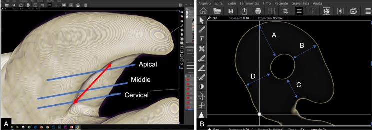
Material and methods: Fourteen extracted maxillary first premolars with a palatal groove on the buccal root were selected. Dentin thickness was measured at 4 stages: initial (M1), after instrumentation (M2), after filling material removal (M3), and after post space preparation (M4). Measurements were taken in 3 root regions: cervical (1 mm coronal to the groove), middle (at the groove's deepest point), and apical (2 mm apical to the groove). Data were analyzed using ANOVA and Tukey's post hoc test (α = 5%).
Results: Significant reductions in dentin thickness were found in all walls across treatment phases. In the palatal wall, dentin thickness dropped below 1 mm after instrumentation, retreatment, and post space preparation. In the buccal and mesial walls, thickness fell below 1 mm after retreatment and/or post space preparation. The distal wall was less affected. The average groove depth was 0.66 ± 0.20 mm, and average groove length was 5.72 ± 1.65 mm.
Conclusions: Post space preparation in maxillary first premolars with palatal groove on the buccal root significantly reduces dentin thickness, especially in the palatal wall, increasing the risk of root weakening. Clinicians should carefully assess the indication of intraradicular posts in such cases to avoid potential complications. Key words:Cone-beam computed tomography, e-vol DX software, Maxillary first premolar, residual dentin thickness, root thickness.
期刊介绍:
Indexed in PUBMED, PubMed Central® (PMC) since 2012 and SCOPUSJournal of Clinical and Experimental Dentistry is an Open Access (free access on-line) - http://www.medicinaoral.com/odo/indice.htm. The aim of the Journal of Clinical and Experimental Dentistry is: - Periodontology - Community and Preventive Dentistry - Esthetic Dentistry - Biomaterials and Bioengineering in Dentistry - Operative Dentistry and Endodontics - Prosthetic Dentistry - Orthodontics - Oral Medicine and Pathology - Odontostomatology for the disabled or special patients - Oral Surgery

 求助内容:
求助内容: 应助结果提醒方式:


