{"title":"Stafne Bone Cavity: A systematic review.","authors":"Paolo Boffano, Muhammad Ruslin","doi":"10.4317/jced.62755","DOIUrl":null,"url":null,"abstract":"<p><strong>Background: </strong>The typical imaging aspect of Stafne Bone Cavities is that of a radiolucent ovoid or round shape image in the posterior mandible below the nerve canal. Anyway, bilobate and bilateral lesions have been reported too, as well as lesions above the inferior alveolar canal. The etiology and pathogenesis are still unknown. The aim of the present article is to review the literature about the current knowledge of this peculiar anatomical condition.</p><p><strong>Material and methods: </strong>The current study was a comprehensive systematic review that was conducted by using databases on the following online sites: PubMed, Embase, SCOPUS, Wiley Online Library, and Ovid MEDLINE.</p><p><strong>Results: </strong>Before applying the filters, 238 publications were identified at first in the considered databases. After the application of the filters, the removal of the duplicates, and the screening process, we ended up with 39 articles that were used in our review. The prevalence of Stafne Bone Cavities oscillates between 0,03% and 3,55%. Mean age ranges between 45,4 and 60,8 years. Males outnumber females, with male:female ratios ranging between 9:4 and 11:0. The most common sites of SBC are observed in the posterior mandible, with body and/or angle regions being the most frequent localization in all studies.</p><p><strong>Discussion: </strong>A wait-and-see approach in terms of a periodic radiograph is recommended in view of the features of this entity, as in exceptional cases tumors seem to have developed in the invaginated salivary gland tissue. <b>Key words:</b>Stafne Bone Cavities, Stafne bone cyst, diagnosis; anatomy, epidemiology.</p>","PeriodicalId":15376,"journal":{"name":"Journal of Clinical and Experimental Dentistry","volume":"17 6","pages":"e725-e731"},"PeriodicalIF":0.0000,"publicationDate":"2025-06-01","publicationTypes":"Journal Article","fieldsOfStudy":null,"isOpenAccess":false,"openAccessPdf":"https://www.ncbi.nlm.nih.gov/pmc/articles/PMC12225759/pdf/","citationCount":"0","resultStr":null,"platform":"Semanticscholar","paperid":null,"PeriodicalName":"Journal of Clinical and Experimental Dentistry","FirstCategoryId":"1085","ListUrlMain":"https://doi.org/10.4317/jced.62755","RegionNum":0,"RegionCategory":null,"ArticlePicture":[],"TitleCN":null,"AbstractTextCN":null,"PMCID":null,"EPubDate":"","PubModel":"","JCR":"Q2","JCRName":"Dentistry","Score":null,"Total":0}
引用次数: 0
Abstract
Background: The typical imaging aspect of Stafne Bone Cavities is that of a radiolucent ovoid or round shape image in the posterior mandible below the nerve canal. Anyway, bilobate and bilateral lesions have been reported too, as well as lesions above the inferior alveolar canal. The etiology and pathogenesis are still unknown. The aim of the present article is to review the literature about the current knowledge of this peculiar anatomical condition.
Material and methods: The current study was a comprehensive systematic review that was conducted by using databases on the following online sites: PubMed, Embase, SCOPUS, Wiley Online Library, and Ovid MEDLINE.
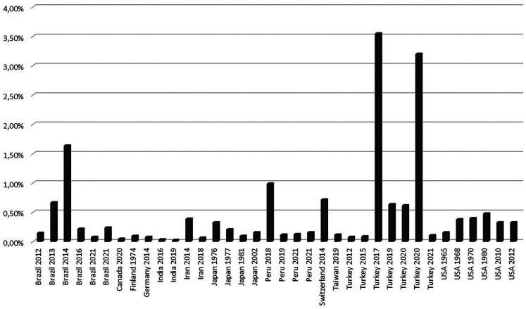
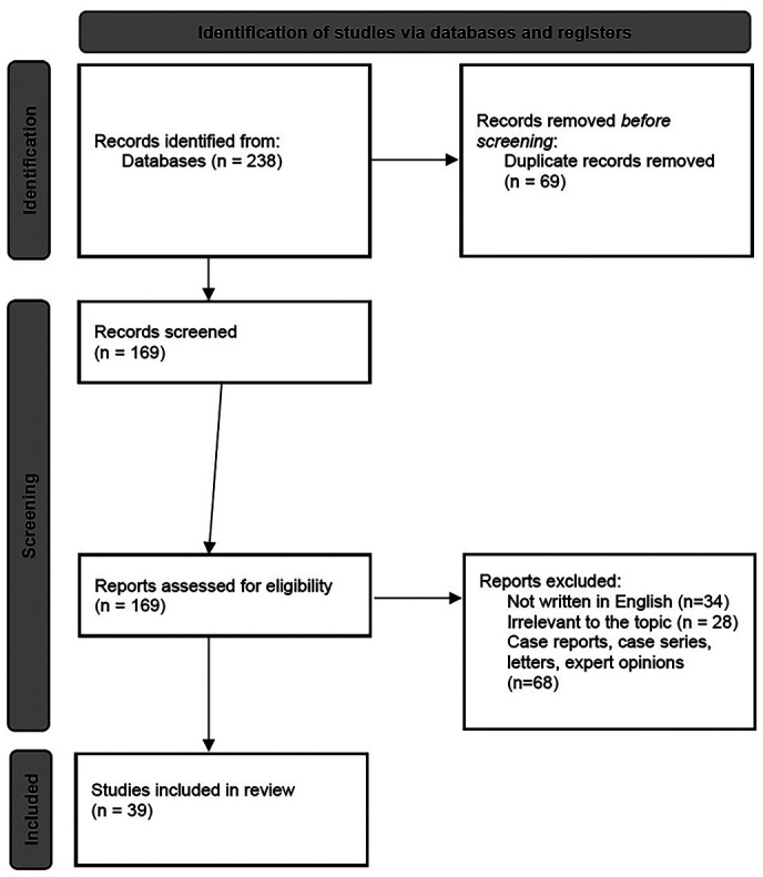
Results: Before applying the filters, 238 publications were identified at first in the considered databases. After the application of the filters, the removal of the duplicates, and the screening process, we ended up with 39 articles that were used in our review. The prevalence of Stafne Bone Cavities oscillates between 0,03% and 3,55%. Mean age ranges between 45,4 and 60,8 years. Males outnumber females, with male:female ratios ranging between 9:4 and 11:0. The most common sites of SBC are observed in the posterior mandible, with body and/or angle regions being the most frequent localization in all studies.
Discussion: A wait-and-see approach in terms of a periodic radiograph is recommended in view of the features of this entity, as in exceptional cases tumors seem to have developed in the invaginated salivary gland tissue. Key words:Stafne Bone Cavities, Stafne bone cyst, diagnosis; anatomy, epidemiology.
期刊介绍:
Indexed in PUBMED, PubMed Central® (PMC) since 2012 and SCOPUSJournal of Clinical and Experimental Dentistry is an Open Access (free access on-line) - http://www.medicinaoral.com/odo/indice.htm. The aim of the Journal of Clinical and Experimental Dentistry is: - Periodontology - Community and Preventive Dentistry - Esthetic Dentistry - Biomaterials and Bioengineering in Dentistry - Operative Dentistry and Endodontics - Prosthetic Dentistry - Orthodontics - Oral Medicine and Pathology - Odontostomatology for the disabled or special patients - Oral Surgery

 求助内容:
求助内容: 应助结果提醒方式:


