Bettina Palicskó, Luca Janovák, László Rejtő, László Váróczy, Zsuzsanna Hevessy, Bettina Kárai
{"title":"Enhancing detection of central nervous system involvement in multiple myeloma: A novel multidimensional dot-plot based analysis for flow cytometry","authors":"Bettina Palicskó, Luca Janovák, László Rejtő, László Váróczy, Zsuzsanna Hevessy, Bettina Kárai","doi":"10.1002/cyto.b.22245","DOIUrl":null,"url":null,"abstract":"<p>Central nervous system (CNS) involvement in multiple myeloma (MM) is a rare but severe complication with a poor prognosis. The identification of malignant plasma cells in cerebrospinal fluid (CSF) is essential for early diagnosis and intervention. However, the sensitivity of traditional diagnostic methods like cytology is low, especially in samples with low-cell counts. This study aimed to develop a multidimensional radar dot-plot analysis using Kaluza software to enhance the sensitivity and specificity of flow cytometry for detecting abnormal plasma cells in CSF. One hundred and twenty-five CSF samples were sent for flow cytometric testing to investigate the central nervous system involvement of MM. Finally, 89 samples from 40 patients were included in our study. Multicolor flow cytometry was performed using an 8-color labeling method, and radar dot-plot analysis was developed using diagnostic bone marrow samples to distinguish normal plasma cells, abnormal plasma cells, and cellular debris. The sensitivity of the novel method was evaluated by diluting myeloma bone marrow cells in pooled CSF samples to simulate low cell counts. Of the 125 CSF specimens, 16 samples from 4 patients showed abnormal plasma cells using both conventional and multidimensional flow cytometry analysis. Discordant results were found in 32 samples (25%), where conventional analysis suggested the presence of abnormal cells, but these were ruled out by multidimensional analysis. Sensitivity testing showed that the multidimensional dot-plot method outperforms conventional two-dimensional dot-plot analysis, as the radar dot plot can be used to identify abnormal cells in samples diluted to 5 WBC/μL, where the cell count of abnormal plasma cells is < 1 cell/μL. Our results showed that the new radar dot-plot analysis can increase the sensitivity and specificity of flow cytometry in MM for the detection of CNS involvement, even in low-cell-count CSF samples, regardless of whether the sample was obtained in a tube containing special reagent or not (TransFix/EDTA CSF Sample Storage tubes). This approach improves diagnostic accuracy, reduces the number of false positive cases caused by antibodies adhering to cell debris, and provides a reliable tool for assessing neurological complications in MM. Further validation is needed in a larger number of cases and testing of the method on different antibody panels.</p>","PeriodicalId":10883,"journal":{"name":"Cytometry Part B: Clinical Cytometry","volume":"108 4","pages":"275-281"},"PeriodicalIF":2.7000,"publicationDate":"2025-07-07","publicationTypes":"Journal Article","fieldsOfStudy":null,"isOpenAccess":false,"openAccessPdf":"https://onlinelibrary.wiley.com/doi/epdf/10.1002/cyto.b.22245","citationCount":"0","resultStr":null,"platform":"Semanticscholar","paperid":null,"PeriodicalName":"Cytometry Part B: Clinical Cytometry","FirstCategoryId":"3","ListUrlMain":"https://onlinelibrary.wiley.com/doi/10.1002/cyto.b.22245","RegionNum":3,"RegionCategory":"医学","ArticlePicture":[],"TitleCN":null,"AbstractTextCN":null,"PMCID":null,"EPubDate":"","PubModel":"","JCR":"Q3","JCRName":"MEDICAL LABORATORY TECHNOLOGY","Score":null,"Total":0}
引用次数: 0
Abstract
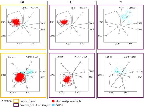
Central nervous system (CNS) involvement in multiple myeloma (MM) is a rare but severe complication with a poor prognosis. The identification of malignant plasma cells in cerebrospinal fluid (CSF) is essential for early diagnosis and intervention. However, the sensitivity of traditional diagnostic methods like cytology is low, especially in samples with low-cell counts. This study aimed to develop a multidimensional radar dot-plot analysis using Kaluza software to enhance the sensitivity and specificity of flow cytometry for detecting abnormal plasma cells in CSF. One hundred and twenty-five CSF samples were sent for flow cytometric testing to investigate the central nervous system involvement of MM. Finally, 89 samples from 40 patients were included in our study. Multicolor flow cytometry was performed using an 8-color labeling method, and radar dot-plot analysis was developed using diagnostic bone marrow samples to distinguish normal plasma cells, abnormal plasma cells, and cellular debris. The sensitivity of the novel method was evaluated by diluting myeloma bone marrow cells in pooled CSF samples to simulate low cell counts. Of the 125 CSF specimens, 16 samples from 4 patients showed abnormal plasma cells using both conventional and multidimensional flow cytometry analysis. Discordant results were found in 32 samples (25%), where conventional analysis suggested the presence of abnormal cells, but these were ruled out by multidimensional analysis. Sensitivity testing showed that the multidimensional dot-plot method outperforms conventional two-dimensional dot-plot analysis, as the radar dot plot can be used to identify abnormal cells in samples diluted to 5 WBC/μL, where the cell count of abnormal plasma cells is < 1 cell/μL. Our results showed that the new radar dot-plot analysis can increase the sensitivity and specificity of flow cytometry in MM for the detection of CNS involvement, even in low-cell-count CSF samples, regardless of whether the sample was obtained in a tube containing special reagent or not (TransFix/EDTA CSF Sample Storage tubes). This approach improves diagnostic accuracy, reduces the number of false positive cases caused by antibodies adhering to cell debris, and provides a reliable tool for assessing neurological complications in MM. Further validation is needed in a larger number of cases and testing of the method on different antibody panels.
期刊介绍:
Cytometry Part B: Clinical Cytometry features original research reports, in-depth reviews and special issues that directly relate to and palpably impact clinical flow, mass and image-based cytometry. These may include clinical and translational investigations important in the diagnostic, prognostic and therapeutic management of patients. Thus, we welcome research papers from various disciplines related [but not limited to] hematopathologists, hematologists, immunologists and cell biologists with clinically relevant and innovative studies investigating individual-cell analytics and/or separations. In addition to the types of papers indicated above, we also welcome Letters to the Editor, describing case reports or important medical or technical topics relevant to our readership without the length and depth of a full original report.
 求助内容:
求助内容: 应助结果提醒方式:


