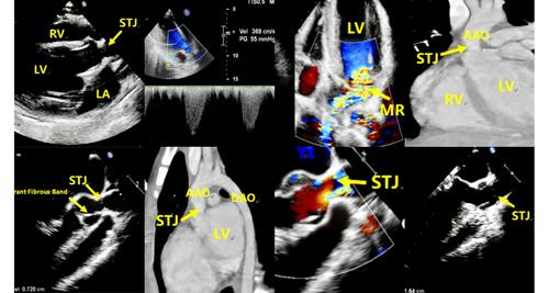
A Rare Cause of Supravalvular Aortic Stenosis: Aberrant Fibrous Band Formation Connecting the Left Coronary Cusp to the Sinus Wall
IF 1.4
4区 医学
Q3 CARDIAC & CARDIOVASCULAR SYSTEMS
Echocardiography-A Journal of Cardiovascular Ultrasound and Allied Techniques
Pub Date : 2025-07-07
DOI:10.1111/echo.70245
引用次数: 0
Abstract
Transesophageal echocardiography (TEE) revealed that supravalvular aortic stenosis was caused by an aberrant fibrous band bridging the left coronary cusp and the sinus wall. This case highlights the advantage of TEE in delineating such abnormal anatomy and guiding precise etiological diagnosis and surgical planning.
瓣上主动脉狭窄的一个罕见原因:连接左冠状动脉尖与窦壁的异常纤维带形成
经食管超声心动图(TEE)显示,瓣上主动脉狭窄是由连接左冠状动脉尖端和窦壁的异常纤维带引起的。本病例强调TEE在描绘此类异常解剖和指导精确病因诊断和手术计划方面的优势。
本文章由计算机程序翻译,如有差异,请以英文原文为准。
求助全文
约1分钟内获得全文
求助全文
来源期刊

CiteScore
2.40
自引率
6.70%
发文量
211
审稿时长
3-6 weeks
期刊介绍:
Echocardiography: A Journal of Cardiovascular Ultrasound and Allied Techniques is the official publication of the International Society of Cardiovascular Ultrasound. Widely recognized for its comprehensive peer-reviewed articles, case studies, original research, and reviews by international authors. Echocardiography keeps its readership of echocardiographers, ultrasound specialists, and cardiologists well informed of the latest developments in the field.
 求助内容:
求助内容: 应助结果提醒方式:


