Anteroposterior displacements of the jaws and incisors relative to the forehead during growth: A longitudinal cephalometric analysis of untreated subjects with Class I occlusions
IF 3.2
Q1 DENTISTRY, ORAL SURGERY & MEDICINE
Journal of the World Federation of Orthodontists
Pub Date : 2025-10-01
DOI:10.1016/j.ejwf.2025.05.006
引用次数: 0
Abstract
Objective
To assess anteroposterior (AP) displacements of the jaws and incisors relative to the forehead in untreated growing subjects with normal (Class I) occlusions.
Materials and Methods
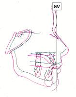
Serial lateral cephalograms from 74 untreated subjects (37 female, 37 male) with Class I dental relationships were obtained from the AAOF Craniofacial Growth Legacy Collection. Mean age at T1 was 9.9 years; T2 was 17.6 years. T1 tracings were secured over millimeter graph paper with Frankfort Horizontal aligned horizontally. Superimpositions were guided by anterior cranial base structures, then shifted along Frankfort Horizontal so that glabella on T1 and T2 shared a common vertical plane (Glabella-Vertical [GV]). AP displacements of Point A (Pt A), Point B (Pt B), maxillary central incisor’s FA point (U1FA), and mandibular central incisor’s FA point (L1FA) were measured relative to GV. Changes in occlusal plane (OP) inclinations were also measured.
Results
Average AP displacement at Pt A was –0.47 mm; Pt B +0.71 mm; U1FA +0.32 mm; and L1FA +0.82 mm. OP inclination decreased 2.30 on average. Displacement of L1FA was significantly different between genders (M, +1.16 mm; F, +0.47 mm; P = 0.04). No significant differences between genders were found for any other variable.
Conclusions
AP jaw and incisor positions were effectively static relative to GV during normal growth between ages 9 and 18 with untreated Class I occlusions. On average, no site underwent AP displacement relative to GV that exceeded 1 mm. The OP tended to rotate counterclockwise. In subjects with Class I dental relationships, pretreatment jaw, and incisor positions do not undergo significant AP changes relative to GV during growth after 9 years of age regardless of gender. GV could thus serve as a reliable AP diagnostic landmark in growing patients.
生长过程中颌骨和门牙相对于前额的前后移位:一项未经治疗的I类咬合受试者的纵向头颅测量分析。
目的:评估正常(I类)牙合未经治疗的生长期受试者的下颌和门牙相对于前额的前后移位。材料和方法:从AAOF颅面生长遗产收藏中获得74名未经治疗的ⅰ类牙关系的受试者(37名女性,37名男性)的连续侧位脑电图。T1时平均年龄9.9岁;T2为17.6岁。T1示踪固定在毫米方格纸上,Frankfort Horizontal水平对齐。在前颅底结构的引导下进行叠加,然后沿法兰克福水平方向移动,使T1和T2的眉间共用一个垂直平面(glabella - vertical [GV])。测量A点(Pt A)、B点(Pt B)、上颌中切牙的FA点(U1FA)、下颌中切牙的FA点(L1FA)相对于GV的AP位移。同时测量了咬合平面(OP)倾斜度的变化。结果:A点AP平均位移-0.47 mm;Pt B +0.71 mm;U1FA +0.32 mm;L1FA +0.82 mm。OP倾角平均下降2.30。L1FA位移在性别间差异显著(M, +1.16 mm;F, +0.47 mm;P = 0.04)。性别之间在其他变量上没有显著差异。结论:在9岁至18岁的正常生长期间,未经治疗的I类咬合,AP颌骨和切牙位置相对于GV有效静止。平均而言,没有部位发生相对于GV超过1mm的AP移位。OP倾向于逆时针旋转。在具有I类牙关系的受试者中,无论性别,预处理颌骨和切牙位置在9岁以后的生长过程中相对于上腭没有明显的AP变化。因此,在生长中的患者中,GV可作为可靠的AP诊断标志。
本文章由计算机程序翻译,如有差异,请以英文原文为准。
求助全文
约1分钟内获得全文
求助全文
来源期刊

Journal of the World Federation of Orthodontists
DENTISTRY, ORAL SURGERY & MEDICINE-
CiteScore
3.80
自引率
4.80%
发文量
34
 求助内容:
求助内容: 应助结果提醒方式:


