{"title":"[Structural changes in the craniofacial complex induced by microimplant-supported skeletal expander - MSE. a review].","authors":"Diana Cecilia Zárate-Guerra, Gissella Gutiérrez-Tapia","doi":"10.21142/2523-2754-1302-2025-243","DOIUrl":null,"url":null,"abstract":"<p><strong>Objective: </strong>The review aimed to assess the structural changes in the craniofacial complex following maxillary expansion assisted by micro-implants, known as the \"<i>Miniscrew assisted rapid palatal expander</i>\" (<i>MARPE</i>) in patients with maxillary transverse deficiency.</p><p><strong>Materials and methods: </strong>The research included a specific type of expander called \"Maxillary Skeletal Expander\" (MSE) the design features a posteriorly positioned screw aligned with the zygomatic buttress, supported by four micro-implants located paramedial to the midpalatal suture with bicortical engagement of the palatal vault and the nasal floor. Four databases (PubMed, Scopus, Embase and ScienceDirect) were reviewed until July 2024. Studies reporting skeletal and dentoalveolar changes in patients with transverse maxillary deficiency evaluated using cone-beam computed tomography (CBCT) were selected. The methodology was ensured using the PRISMA checklist.</p><p><strong>Results: </strong>13 articles were selected according to the inclusion criteria. Regarding skeletal changes, the disjunction in the axial cut is parallel or nearly parallel, both in lineal and angular measurements (the posterior nasal spine represents between 82% and 101% in relation to anterior nasal spine) Two studies report a deviation in the disjunction at the ANS level of 1.1mm (+/- 1mm) in half of the sample, between one side and the other. Parallelism depends on the disarticulation of the pterygopalatine suture. A correlation was reported between bicortical anchorage anc the opening of this suture (P=0.0003) in 55.6% of the evaluated patients. In the coronal cut the expansion is pyramidal with an upper vertex, and the fulcrum would be located at the most external and inferior point of the frontozygomatic suture, allowing a rotation of 0.6° of the zygomaticomaxillary complex for each millimeter of expansion. Regarding dentoalveolar changes, studies find an average buccal inclination of the upper first molars of 3°.</p><p><strong>Conclusions: </strong>Maxillary expansion assisted by micro-implants significantly impacts craniofacial structures. Furthermore, by ensuring orthopedic movements in young and young adult patients, adverse dental and periodontal effects associated with compensatory or non-skeletal expansion are reduced. Further studies on larger populations are required to confirm these effects.</p>","PeriodicalId":33326,"journal":{"name":"Revista Cientifica Odontologica","volume":"13 2","pages":"e243"},"PeriodicalIF":0.0000,"publicationDate":"2025-05-16","publicationTypes":"Journal Article","fieldsOfStudy":null,"isOpenAccess":false,"openAccessPdf":"https://www.ncbi.nlm.nih.gov/pmc/articles/PMC12217047/pdf/","citationCount":"0","resultStr":null,"platform":"Semanticscholar","paperid":null,"PeriodicalName":"Revista Cientifica Odontologica","FirstCategoryId":"1085","ListUrlMain":"https://doi.org/10.21142/2523-2754-1302-2025-243","RegionNum":0,"RegionCategory":null,"ArticlePicture":[],"TitleCN":null,"AbstractTextCN":null,"PMCID":null,"EPubDate":"2025/4/1 0:00:00","PubModel":"eCollection","JCR":"","JCRName":"","Score":null,"Total":0}
引用次数: 0
Abstract
Objective: The review aimed to assess the structural changes in the craniofacial complex following maxillary expansion assisted by micro-implants, known as the "Miniscrew assisted rapid palatal expander" (MARPE) in patients with maxillary transverse deficiency.
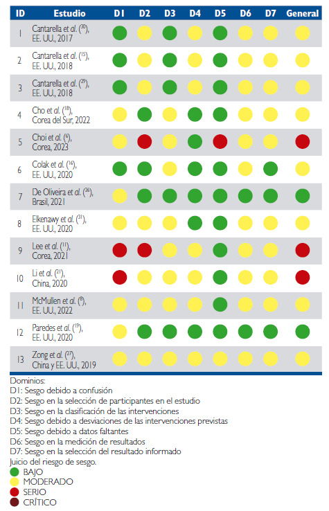
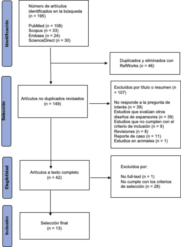
Materials and methods: The research included a specific type of expander called "Maxillary Skeletal Expander" (MSE) the design features a posteriorly positioned screw aligned with the zygomatic buttress, supported by four micro-implants located paramedial to the midpalatal suture with bicortical engagement of the palatal vault and the nasal floor. Four databases (PubMed, Scopus, Embase and ScienceDirect) were reviewed until July 2024. Studies reporting skeletal and dentoalveolar changes in patients with transverse maxillary deficiency evaluated using cone-beam computed tomography (CBCT) were selected. The methodology was ensured using the PRISMA checklist.
Results: 13 articles were selected according to the inclusion criteria. Regarding skeletal changes, the disjunction in the axial cut is parallel or nearly parallel, both in lineal and angular measurements (the posterior nasal spine represents between 82% and 101% in relation to anterior nasal spine) Two studies report a deviation in the disjunction at the ANS level of 1.1mm (+/- 1mm) in half of the sample, between one side and the other. Parallelism depends on the disarticulation of the pterygopalatine suture. A correlation was reported between bicortical anchorage anc the opening of this suture (P=0.0003) in 55.6% of the evaluated patients. In the coronal cut the expansion is pyramidal with an upper vertex, and the fulcrum would be located at the most external and inferior point of the frontozygomatic suture, allowing a rotation of 0.6° of the zygomaticomaxillary complex for each millimeter of expansion. Regarding dentoalveolar changes, studies find an average buccal inclination of the upper first molars of 3°.
Conclusions: Maxillary expansion assisted by micro-implants significantly impacts craniofacial structures. Furthermore, by ensuring orthopedic movements in young and young adult patients, adverse dental and periodontal effects associated with compensatory or non-skeletal expansion are reduced. Further studies on larger populations are required to confirm these effects.

 求助内容:
求助内容: 应助结果提醒方式:


