{"title":"Evaluation of Enamel Surface Roughness and Volumetric Change after Resin Remnant Removal following Orthodontic Bracket Debonding.","authors":"Bora Korkut, Kadir Emre Uzun, Cigdem Hacıali, Tuna Unal, Dilek Tagtekin","doi":"10.3290/j.ohpd.c_2117","DOIUrl":null,"url":null,"abstract":"<p><strong>Purpose: </strong>To evaluate surface roughness and volumetric change of enamel after using different resin remnant removal (RRR) techniques, following orthodontic bracket debonding.</p><p><strong>Materials and methods: </strong>Metal orthodontic brackets (Mini Twin Brackets, RMO) were bonded to 60 human (central or lateral) labial mid-third surfaces, and debonded 24 h after by a single orthodontist. The remaining composites were completely removed with the fluorescence light guidance by the D-Light-Pro led curing unit (GC/detection mode). The removal procedures were performed without magnification (n = 30) or with 20× magnification/5500 K illumination by a dental microscope (OMS2000, Zumax) (n = 30). Three RRR techniques were used: 12-bladed carbide bur (Horico), red-banded diamond bur (Horico), SofLex Disc (medium/40 μm, fine/24 μm, and superfine/8 µm; 3M). Surface changes were evaluated visually through microscope photographs by enamel surface index (ESI) and volumetrically by overlapping the three-dimensional images of a laser scanner device (LAS-20, SD-Mechatronik) in the Geomagic Design X (3D Systems) software. The deemed significance was set at 0.050 for the statistical analyses.</p><p><strong>Results: </strong>A positive, strong correlation was found between visual and volumetric change scores (P 0.001). Lesser volumetric loss (P 0.001) and roughness (P = 0.009) were observed for all RRR techniques when the magnification was used. Volumetric loss (mm3) by diamond bur was significantly the highest [1.85(1-3)a], followed by SofLex Disc [1.1(1-1)c] and carbide bur [0.59(0-1)b](P 0.001). Visual surface roughness scores (Ra) were significantly higher for diamond bur [4.5(4-5)b](P 0.001), followed by carbide bur 2(1-3)a and SofLex Disc 1(1-2)a.</p><p><strong>Conclusion: </strong>Surface roughness should always be assessed together with the volumetric enamel loss for the selection of RRR technique. Red-banded diamond bur should not be used for RRR. Even though the least surface roughness can be provided by SofLex Disc system, it can provide more intact enamel surface loss than the carbide bur. Magnification was considered useful for the RRR to provide a smoother surface while better preserving the intact enamel tissue.</p>","PeriodicalId":19696,"journal":{"name":"Oral health & preventive dentistry","volume":"23 ","pages":"355-364"},"PeriodicalIF":1.4000,"publicationDate":"2025-07-04","publicationTypes":"Journal Article","fieldsOfStudy":null,"isOpenAccess":false,"openAccessPdf":"https://www.ncbi.nlm.nih.gov/pmc/articles/PMC12246803/pdf/","citationCount":"0","resultStr":null,"platform":"Semanticscholar","paperid":null,"PeriodicalName":"Oral health & preventive dentistry","FirstCategoryId":"3","ListUrlMain":"https://doi.org/10.3290/j.ohpd.c_2117","RegionNum":4,"RegionCategory":"医学","ArticlePicture":[],"TitleCN":null,"AbstractTextCN":null,"PMCID":null,"EPubDate":"","PubModel":"","JCR":"Q3","JCRName":"DENTISTRY, ORAL SURGERY & MEDICINE","Score":null,"Total":0}
引用次数: 0
Abstract
Purpose: To evaluate surface roughness and volumetric change of enamel after using different resin remnant removal (RRR) techniques, following orthodontic bracket debonding.
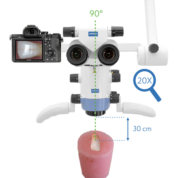
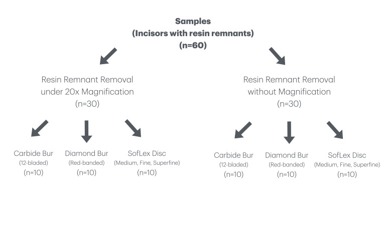
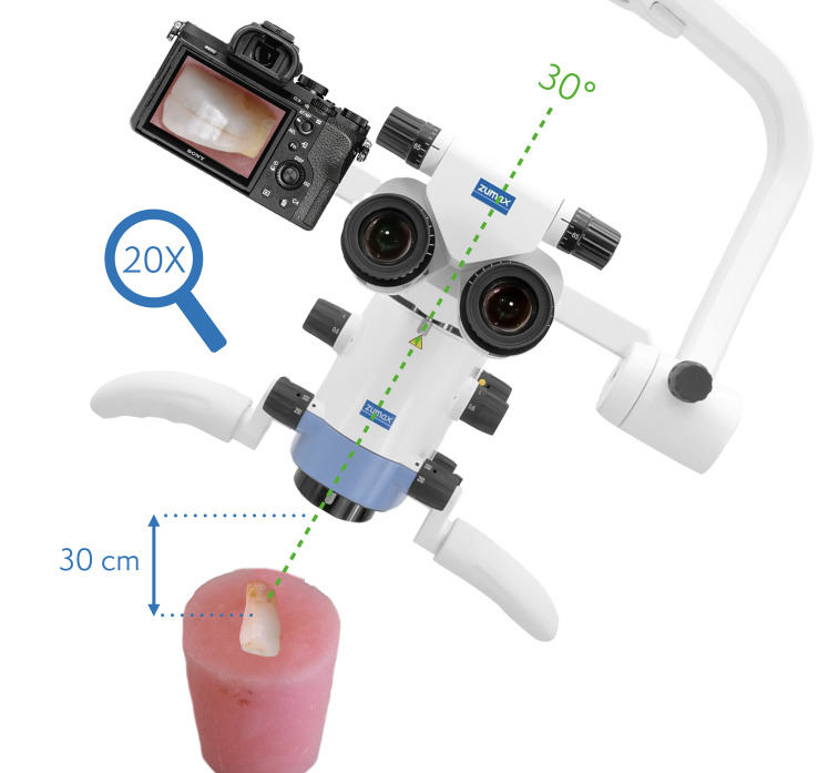
Materials and methods: Metal orthodontic brackets (Mini Twin Brackets, RMO) were bonded to 60 human (central or lateral) labial mid-third surfaces, and debonded 24 h after by a single orthodontist. The remaining composites were completely removed with the fluorescence light guidance by the D-Light-Pro led curing unit (GC/detection mode). The removal procedures were performed without magnification (n = 30) or with 20× magnification/5500 K illumination by a dental microscope (OMS2000, Zumax) (n = 30). Three RRR techniques were used: 12-bladed carbide bur (Horico), red-banded diamond bur (Horico), SofLex Disc (medium/40 μm, fine/24 μm, and superfine/8 µm; 3M). Surface changes were evaluated visually through microscope photographs by enamel surface index (ESI) and volumetrically by overlapping the three-dimensional images of a laser scanner device (LAS-20, SD-Mechatronik) in the Geomagic Design X (3D Systems) software. The deemed significance was set at 0.050 for the statistical analyses.
Results: A positive, strong correlation was found between visual and volumetric change scores (P 0.001). Lesser volumetric loss (P 0.001) and roughness (P = 0.009) were observed for all RRR techniques when the magnification was used. Volumetric loss (mm3) by diamond bur was significantly the highest [1.85(1-3)a], followed by SofLex Disc [1.1(1-1)c] and carbide bur [0.59(0-1)b](P 0.001). Visual surface roughness scores (Ra) were significantly higher for diamond bur [4.5(4-5)b](P 0.001), followed by carbide bur 2(1-3)a and SofLex Disc 1(1-2)a.
Conclusion: Surface roughness should always be assessed together with the volumetric enamel loss for the selection of RRR technique. Red-banded diamond bur should not be used for RRR. Even though the least surface roughness can be provided by SofLex Disc system, it can provide more intact enamel surface loss than the carbide bur. Magnification was considered useful for the RRR to provide a smoother surface while better preserving the intact enamel tissue.
期刊介绍:
Clinicians, general practitioners, teachers, researchers, and public health administrators will find this journal an indispensable source of essential, timely information about scientific progress in the fields of oral health and the prevention of caries, periodontal diseases, oral mucosal diseases, and dental trauma. Central topics, including oral hygiene, oral epidemiology, oral health promotion, and public health issues, are covered in peer-reviewed articles such as clinical and basic science research reports; reviews; invited focus articles, commentaries, and guest editorials; and symposium, workshop, and conference proceedings.
 求助内容:
求助内容: 应助结果提醒方式:


