{"title":"Shared neural signatures of photophobia in migraine and post-traumatic headache: a task-based fMRI study.","authors":"Rune Häckert Christensen, Haidar Muhsen Al-Khazali, Anna Gudrun Melchior, Messoud Ashina, Håkan Ashina","doi":"10.1186/s10194-025-02088-y","DOIUrl":null,"url":null,"abstract":"<p><strong>Background: </strong>Persistent post-traumatic headache (PTH) and migraine frequently present with photic hypersensitivity that exacerbates headache symptoms. We sought to determine whether persistent PTH is associated with altered brain responses to visual stimuli and to explore shared neural mechanisms of photophobia with migraine.</p><p><strong>Methods: </strong>This cross-sectional functional magnetic resonance imaging (fMRI) study included 80 adults with persistent PTH, 261 with migraine, and 143 healthy controls (HCs). All participants underwent visual stimulation using a flickering checkerboard during a 3T fMRI session. Blood oxygen level-dependent (BOLD) responses were examined using whole-brain and region-of-interest (ROI) analyses. All analyses were adjusted for age and sex.</p><p><strong>Results: </strong>Whole-brain analysis revealed no significant BOLD differences across the full persistent PTH, migraine, and HC groups. However, participants with persistent PTH who experienced photophobia during the scan (n = 41) showed greater activation in the anterior and midcingulate cortex compared with HCs (P<sub>FWE</sub> = 0.010). No differences were observed between photophobic participants with persistent PTH and those with migraine who reported an attack during the fMRI session. ROI analyses identified greater activation in the anterior cingulate, midcingulate, and insular cortices in both photophobic participants with persistent PTH and ictal participants with migraine, relative to HCs (all P < 0.05). No significant differences were found between photophobic participants with persistent PTH and ictal participants with migraine.</p><p><strong>Conclusions: </strong>Photophobia in persistent PTH is associated with greater activation in cortical regions implicated in pain processing. These patterns parallel those observed during migraine attacks, indicating shared neural mechanisms between the two headache disorders.</p>","PeriodicalId":16013,"journal":{"name":"Journal of Headache and Pain","volume":"26 1","pages":"154"},"PeriodicalIF":7.9000,"publicationDate":"2025-07-03","publicationTypes":"Journal Article","fieldsOfStudy":null,"isOpenAccess":false,"openAccessPdf":"https://www.ncbi.nlm.nih.gov/pmc/articles/PMC12231636/pdf/","citationCount":"0","resultStr":null,"platform":"Semanticscholar","paperid":null,"PeriodicalName":"Journal of Headache and Pain","FirstCategoryId":"3","ListUrlMain":"https://doi.org/10.1186/s10194-025-02088-y","RegionNum":1,"RegionCategory":"医学","ArticlePicture":[],"TitleCN":null,"AbstractTextCN":null,"PMCID":null,"EPubDate":"","PubModel":"","JCR":"Q1","JCRName":"CLINICAL NEUROLOGY","Score":null,"Total":0}
引用次数: 0
Abstract
Background: Persistent post-traumatic headache (PTH) and migraine frequently present with photic hypersensitivity that exacerbates headache symptoms. We sought to determine whether persistent PTH is associated with altered brain responses to visual stimuli and to explore shared neural mechanisms of photophobia with migraine.
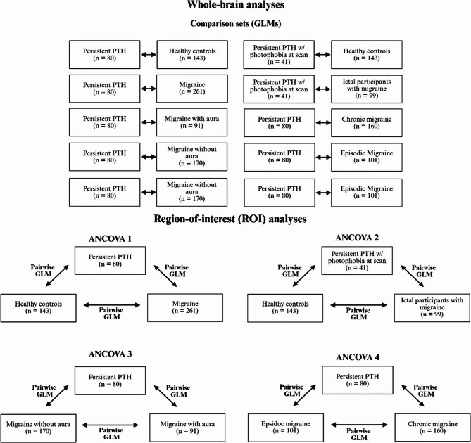
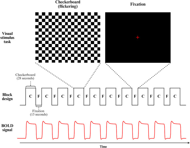
Methods: This cross-sectional functional magnetic resonance imaging (fMRI) study included 80 adults with persistent PTH, 261 with migraine, and 143 healthy controls (HCs). All participants underwent visual stimulation using a flickering checkerboard during a 3T fMRI session. Blood oxygen level-dependent (BOLD) responses were examined using whole-brain and region-of-interest (ROI) analyses. All analyses were adjusted for age and sex.
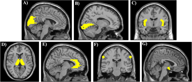
Results: Whole-brain analysis revealed no significant BOLD differences across the full persistent PTH, migraine, and HC groups. However, participants with persistent PTH who experienced photophobia during the scan (n = 41) showed greater activation in the anterior and midcingulate cortex compared with HCs (PFWE = 0.010). No differences were observed between photophobic participants with persistent PTH and those with migraine who reported an attack during the fMRI session. ROI analyses identified greater activation in the anterior cingulate, midcingulate, and insular cortices in both photophobic participants with persistent PTH and ictal participants with migraine, relative to HCs (all P < 0.05). No significant differences were found between photophobic participants with persistent PTH and ictal participants with migraine.
Conclusions: Photophobia in persistent PTH is associated with greater activation in cortical regions implicated in pain processing. These patterns parallel those observed during migraine attacks, indicating shared neural mechanisms between the two headache disorders.
期刊介绍:
The Journal of Headache and Pain, a peer-reviewed open-access journal published under the BMC brand, a part of Springer Nature, is dedicated to researchers engaged in all facets of headache and related pain syndromes. It encompasses epidemiology, public health, basic science, translational medicine, clinical trials, and real-world data.
With a multidisciplinary approach, The Journal of Headache and Pain addresses headache medicine and related pain syndromes across all medical disciplines. It particularly encourages submissions in clinical, translational, and basic science fields, focusing on pain management, genetics, neurology, and internal medicine. The journal publishes research articles, reviews, letters to the Editor, as well as consensus articles and guidelines, aimed at promoting best practices in managing patients with headaches and related pain.
 求助内容:
求助内容: 应助结果提醒方式:


