Xiaoyu Jiang, Mary Kay Washington, Manhal J Izzy, Gracie Piantek, Ming Lu, Xinqiang Yan, John C Gore, Junzhong Xu
{"title":"Noninvasive assessment of liver inflammation in metabolic dysfunction associated steatohepatitis using MR cytometry.","authors":"Xiaoyu Jiang, Mary Kay Washington, Manhal J Izzy, Gracie Piantek, Ming Lu, Xinqiang Yan, John C Gore, Junzhong Xu","doi":"10.1038/s44303-025-00080-4","DOIUrl":null,"url":null,"abstract":"<p><p>The current diagnostic gold standard for metabolic dysfunction-associated steatohepatitis (MASH) requires invasive biopsy to assess steatosis, inflammation, and ballooning. While MRI-based proton density fat fraction (PDFF) and MR elastography address steatosis and fibrosis, non-invasive methods for evaluating hepatic inflammation remain lacking. This study developed a diffusion MRI (dMRI)-based MR cytometry technique to map liver cellular properties, including MRI-derived cell size (excluding fat content) and cell density. Validation through histology-driven simulations and ex vivo MRI of fixed human liver specimens demonstrated that stromal regions exhibit smaller MRI-derived cell sizes and higher cell densities than both normal and fatty tissues. An in vivo feasibility study, conducted on healthy subjects (n = 5) and MASH patients (n = 5) using a clinical 3 T MRI system, further showcased the potential of MR cytometry to characterize pathological changes in liver microstructure.</p>","PeriodicalId":501709,"journal":{"name":"npj Imaging","volume":"3 1","pages":"17"},"PeriodicalIF":0.0000,"publicationDate":"2025-05-03","publicationTypes":"Journal Article","fieldsOfStudy":null,"isOpenAccess":false,"openAccessPdf":"https://www.ncbi.nlm.nih.gov/pmc/articles/PMC12118690/pdf/","citationCount":"0","resultStr":null,"platform":"Semanticscholar","paperid":null,"PeriodicalName":"npj Imaging","FirstCategoryId":"1085","ListUrlMain":"https://doi.org/10.1038/s44303-025-00080-4","RegionNum":0,"RegionCategory":null,"ArticlePicture":[],"TitleCN":null,"AbstractTextCN":null,"PMCID":null,"EPubDate":"","PubModel":"","JCR":"","JCRName":"","Score":null,"Total":0}
引用次数: 0
Abstract
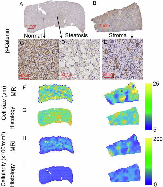
The current diagnostic gold standard for metabolic dysfunction-associated steatohepatitis (MASH) requires invasive biopsy to assess steatosis, inflammation, and ballooning. While MRI-based proton density fat fraction (PDFF) and MR elastography address steatosis and fibrosis, non-invasive methods for evaluating hepatic inflammation remain lacking. This study developed a diffusion MRI (dMRI)-based MR cytometry technique to map liver cellular properties, including MRI-derived cell size (excluding fat content) and cell density. Validation through histology-driven simulations and ex vivo MRI of fixed human liver specimens demonstrated that stromal regions exhibit smaller MRI-derived cell sizes and higher cell densities than both normal and fatty tissues. An in vivo feasibility study, conducted on healthy subjects (n = 5) and MASH patients (n = 5) using a clinical 3 T MRI system, further showcased the potential of MR cytometry to characterize pathological changes in liver microstructure.

 求助内容:
求助内容: 应助结果提醒方式:


