Radiographic relationship of third molars with the mandibular canal as a predictor of inferior alveolar nerve sensory disturbance: A systematic review and meta-analysis.
Abbas Shokri, Ashkan Sadeghi Farnia, Ali Heidari, Forough Abbasiyan, Behnaz Alafchi
{"title":"Radiographic relationship of third molars with the mandibular canal as a predictor of inferior alveolar nerve sensory disturbance: A systematic review and meta-analysis.","authors":"Abbas Shokri, Ashkan Sadeghi Farnia, Ali Heidari, Forough Abbasiyan, Behnaz Alafchi","doi":"10.5624/isd.20240243","DOIUrl":null,"url":null,"abstract":"<p><strong>Purpose: </strong>This study was performed to assess the relationship of the third molars with the mandibular canal as a predictor of inferior alveolar nerve (IAN) sensory disturbances using panoramic radiography (PR) and cone-beam computed tomography (CBCT).</p><p><strong>Materials and methods: </strong>A systematic search was conducted of 4 databases-PubMed, Scopus, Web of Science, and Google Scholar-for the period from 1985 to 2024. In the retrieved articles, the outcome of interest was the relationship of the mandibular canal with the third molars on PR and CBCT scans. The risk of bias was assessed using the Newcastle-Ottawa Scale, and quantitative meta-analysis was performed using STATA. A random-effects restricted maximum likelihood model was employed for the meta-analysis, and the I<sup>2</sup> statistic was used to assess heterogeneity.</p><p><strong>Results: </strong>A total of 1,635 articles were initially retrieved. After a rigorous selection process, 20 studies were included in the qualitative synthesis, and 8 were selected for the meta-analysis. The findings indicated that CBCT yielded higher prevalence rates for root darkening, root deflection, interruption of the white line, diversion of the mandibular canal, and narrowing of the mandibular canal (theta values: 49.962, 4.76, 8.09, 2.229, and 4.708, respectively) compared with PR (theta values: 1.363, 1.605, 6.322, 0.655, and 1.449, respectively).</p><p><strong>Conclusion: </strong>CBCT was more accurate than PR in investigating predictors of IAN paresthesia in mandibular third molar surgery. Considering the higher prevalence of paresthesia in the presence of root darkening, CBCT may be highly efficient in detecting this parameter and thus aiding in the prevention of paresthesia.</p>","PeriodicalId":51714,"journal":{"name":"Imaging Science in Dentistry","volume":"55 2","pages":"114-125"},"PeriodicalIF":2.1000,"publicationDate":"2025-06-01","publicationTypes":"Journal Article","fieldsOfStudy":null,"isOpenAccess":false,"openAccessPdf":"https://www.ncbi.nlm.nih.gov/pmc/articles/PMC12210119/pdf/","citationCount":"0","resultStr":null,"platform":"Semanticscholar","paperid":null,"PeriodicalName":"Imaging Science in Dentistry","FirstCategoryId":"1085","ListUrlMain":"https://doi.org/10.5624/isd.20240243","RegionNum":0,"RegionCategory":null,"ArticlePicture":[],"TitleCN":null,"AbstractTextCN":null,"PMCID":null,"EPubDate":"2025/4/28 0:00:00","PubModel":"Epub","JCR":"Q3","JCRName":"DENTISTRY, ORAL SURGERY & MEDICINE","Score":null,"Total":0}
引用次数: 0
Abstract
Purpose: This study was performed to assess the relationship of the third molars with the mandibular canal as a predictor of inferior alveolar nerve (IAN) sensory disturbances using panoramic radiography (PR) and cone-beam computed tomography (CBCT).
Materials and methods: A systematic search was conducted of 4 databases-PubMed, Scopus, Web of Science, and Google Scholar-for the period from 1985 to 2024. In the retrieved articles, the outcome of interest was the relationship of the mandibular canal with the third molars on PR and CBCT scans. The risk of bias was assessed using the Newcastle-Ottawa Scale, and quantitative meta-analysis was performed using STATA. A random-effects restricted maximum likelihood model was employed for the meta-analysis, and the I2 statistic was used to assess heterogeneity.
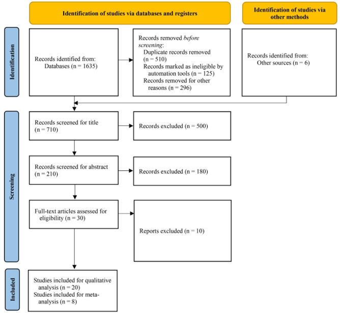
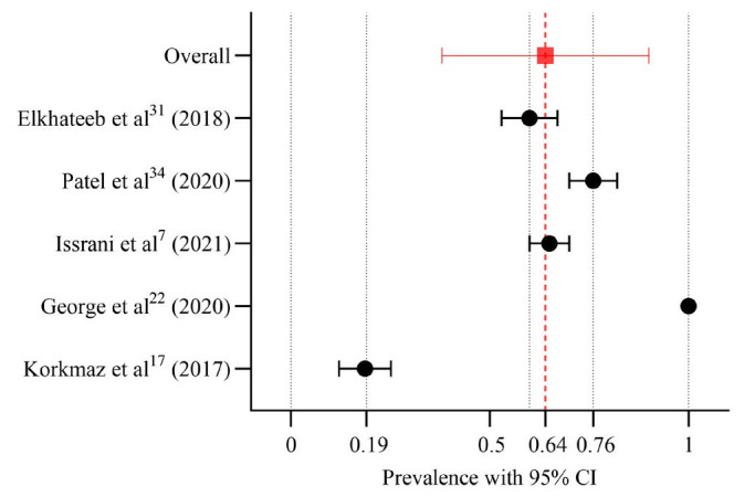
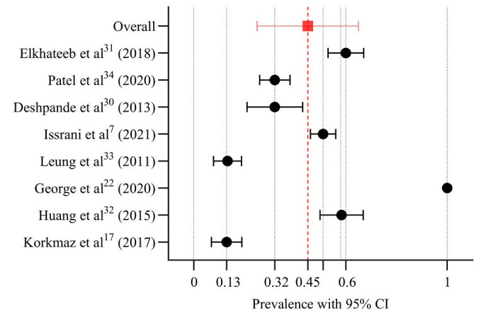
Results: A total of 1,635 articles were initially retrieved. After a rigorous selection process, 20 studies were included in the qualitative synthesis, and 8 were selected for the meta-analysis. The findings indicated that CBCT yielded higher prevalence rates for root darkening, root deflection, interruption of the white line, diversion of the mandibular canal, and narrowing of the mandibular canal (theta values: 49.962, 4.76, 8.09, 2.229, and 4.708, respectively) compared with PR (theta values: 1.363, 1.605, 6.322, 0.655, and 1.449, respectively).
Conclusion: CBCT was more accurate than PR in investigating predictors of IAN paresthesia in mandibular third molar surgery. Considering the higher prevalence of paresthesia in the presence of root darkening, CBCT may be highly efficient in detecting this parameter and thus aiding in the prevention of paresthesia.

 求助内容:
求助内容: 应助结果提醒方式:


