Diagnostic performance of deep learning models in classifying mandibular third molar and mandibular canal contact status on panoramic radiographs: A systematic review and meta-analysis.
{"title":"Diagnostic performance of deep learning models in classifying mandibular third molar and mandibular canal contact status on panoramic radiographs: A systematic review and meta-analysis.","authors":"Hamza Al Salieti, Hala Al Sliti, Saleh Alkadi","doi":"10.5624/isd.20240239","DOIUrl":null,"url":null,"abstract":"<p><strong>Purpose: </strong>Panoramic radiographs have recently become a platform for deep learning models, which show potential in enhancing diagnostic accuracy for detecting contact between mandibular third molars and the mandibular canal. However, detailed information regarding the accuracy of these models in identifying such contact remains limited.</p><p><strong>Materials and methods: </strong>In accordance with the PRISMA-2020 and PRISMA-DTA guidelines, the PubMed, ScienceDirect, Web of Science, Embase, and EBSCO databases were systematically searched up to September 2024. Eligible studies employed deep learning models based on convolutional neural networks to classify the contact between mandibular third molars and the mandibular canal. Extracted metrics included accuracy, sensitivity, specificity, precision, and F1-score. A meta-analysis using random effects models pooled these performance metrics, while univariate and multivariate meta-regressions were conducted to explore sources of heterogeneity. Study quality was assessed using the QUADAS-2 tool.</p><p><strong>Results: </strong>Seven studies incorporating 4,955 panoramic radiographs reported pooled performance metrics of 83.4% accuracy, 80.2% sensitivity, 85.8% specificity, 83.3% precision, and an F1-score of 80.9%. High heterogeneity (I<sup>2</sup> > 90%) was primarily attributable to variations in sample size, image resolution, model architecture, and model complexity. Meta-regression analyses identified image resolution and architecture (e.g., VGG-16, AlexNet) as key factors. Although the overall risk of bias was low, the patient selection domain was often unclear.</p><p><strong>Conclusion: </strong>Deep learning models exhibit significant promise in evaluating mandibular third molar and mandibular canal contact on panoramic radiographs, potentially complementing traditional methods. The adoption of standardized protocols, diverse datasets, and explainable artificial intelligence will be crucial for broader clinical application.</p>","PeriodicalId":51714,"journal":{"name":"Imaging Science in Dentistry","volume":"55 2","pages":"139-150"},"PeriodicalIF":2.1000,"publicationDate":"2025-06-01","publicationTypes":"Journal Article","fieldsOfStudy":null,"isOpenAccess":false,"openAccessPdf":"https://www.ncbi.nlm.nih.gov/pmc/articles/PMC12210121/pdf/","citationCount":"0","resultStr":null,"platform":"Semanticscholar","paperid":null,"PeriodicalName":"Imaging Science in Dentistry","FirstCategoryId":"1085","ListUrlMain":"https://doi.org/10.5624/isd.20240239","RegionNum":0,"RegionCategory":null,"ArticlePicture":[],"TitleCN":null,"AbstractTextCN":null,"PMCID":null,"EPubDate":"2025/4/10 0:00:00","PubModel":"Epub","JCR":"Q3","JCRName":"DENTISTRY, ORAL SURGERY & MEDICINE","Score":null,"Total":0}
引用次数: 0
Abstract
Purpose: Panoramic radiographs have recently become a platform for deep learning models, which show potential in enhancing diagnostic accuracy for detecting contact between mandibular third molars and the mandibular canal. However, detailed information regarding the accuracy of these models in identifying such contact remains limited.
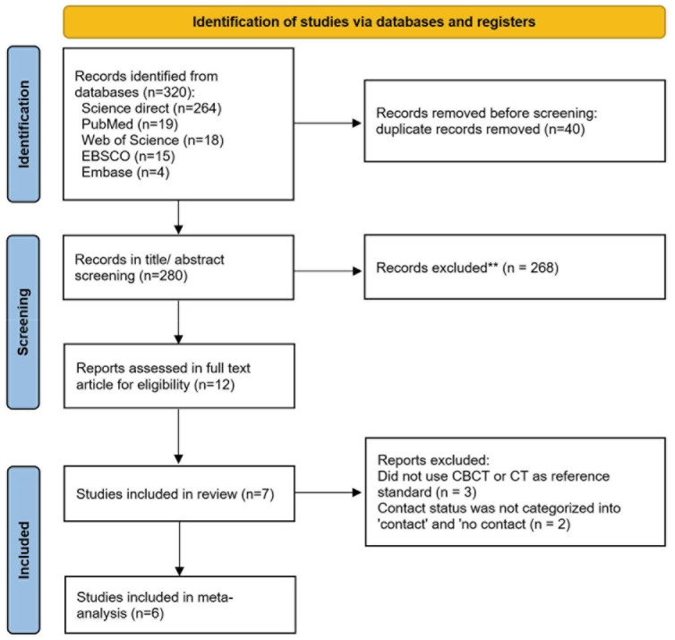
Materials and methods: In accordance with the PRISMA-2020 and PRISMA-DTA guidelines, the PubMed, ScienceDirect, Web of Science, Embase, and EBSCO databases were systematically searched up to September 2024. Eligible studies employed deep learning models based on convolutional neural networks to classify the contact between mandibular third molars and the mandibular canal. Extracted metrics included accuracy, sensitivity, specificity, precision, and F1-score. A meta-analysis using random effects models pooled these performance metrics, while univariate and multivariate meta-regressions were conducted to explore sources of heterogeneity. Study quality was assessed using the QUADAS-2 tool.
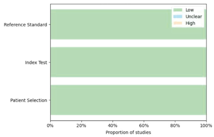
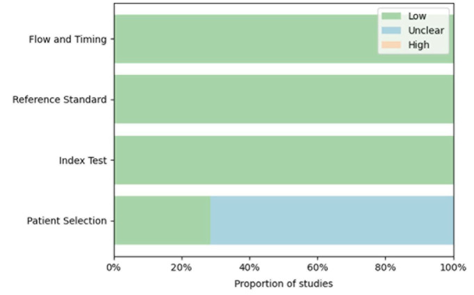
Results: Seven studies incorporating 4,955 panoramic radiographs reported pooled performance metrics of 83.4% accuracy, 80.2% sensitivity, 85.8% specificity, 83.3% precision, and an F1-score of 80.9%. High heterogeneity (I2 > 90%) was primarily attributable to variations in sample size, image resolution, model architecture, and model complexity. Meta-regression analyses identified image resolution and architecture (e.g., VGG-16, AlexNet) as key factors. Although the overall risk of bias was low, the patient selection domain was often unclear.
Conclusion: Deep learning models exhibit significant promise in evaluating mandibular third molar and mandibular canal contact on panoramic radiographs, potentially complementing traditional methods. The adoption of standardized protocols, diverse datasets, and explainable artificial intelligence will be crucial for broader clinical application.
目的:全景x线照片最近成为深度学习模型的一个平台,它在检测下颌第三磨牙与下颌管之间的接触方面显示出提高诊断准确性的潜力。然而,关于这些模型在确定这种接触方面的准确性的详细信息仍然有限。材料和方法:根据PRISMA-2020和PRISMA-DTA指南,系统检索了PubMed、ScienceDirect、Web of Science、Embase和EBSCO数据库,检索时间截止到2024年9月。符合条件的研究采用基于卷积神经网络的深度学习模型对下颌第三磨牙与下颌管之间的接触进行分类。提取的指标包括准确性、敏感性、特异性、精密度和f1评分。使用随机效应模型的荟萃分析汇集了这些绩效指标,同时进行单变量和多变量荟萃回归来探索异质性的来源。使用QUADAS-2工具评估研究质量。结果:包含4,955张全景x线片的7项研究报告了83.4%的准确率、80.2%的灵敏度、85.8%的特异性、83.3%的精确度和80.9%的f1评分。高异质性(90%)主要归因于样本大小、图像分辨率、模型架构和模型复杂性的差异。元回归分析确定图像分辨率和架构(例如,VGG-16, AlexNet)是关键因素。尽管总体偏倚风险较低,但患者选择领域往往不明确。结论:深度学习模型在评估下颌第三磨牙和下颌管在全景x线片上的接触方面具有重要的前景,可能是传统方法的补充。采用标准化的方案、多样化的数据集和可解释的人工智能对于更广泛的临床应用至关重要。

 求助内容:
求助内容: 应助结果提醒方式:


