{"title":"Retrospective analysis of computed tomography examinations in patients with lower abdominal pain: A single-center experience.","authors":"Kouichi Asahi","doi":"10.4329/wjr.v17.i6.105632","DOIUrl":null,"url":null,"abstract":"<p><strong>Background: </strong>Lower abdominal pain presents as a common complaint with diverse etiologies ranging from benign conditions to surgical emergencies. Computed tomography (CT) imaging has become instrumental in accurate diagnosis and management planning, though clinicians must carefully balance its benefits against radiation exposure concerns and resource allocation constraints.</p><p><strong>Aim: </strong>To comprehensively evaluate the diagnostic accuracy, utility and clinical implications of CT in patients with acute lower abdominal pain, emphasizing rare anatomical variations, pregnancy-related issues, and practical limitations of other imaging modalities in emergency settings in Japan.</p><p><strong>Methods: </strong>This retrospective review included 230 patients who underwent CT scans for acute lower abdominal pain at Juntendo University Shizuoka Hospital throughout 2014. CT findings were systematically correlated with the final clinical diagnoses. Cases with diagnostic uncertain underwent independent reviewed by two experienced radiologists. The CT protocols included portal venous-phase imaging with selective arterial-phase acquisition when clinically indicated.</p><p><strong>Results: </strong>Idiopathic pain was the most common diagnosis (104 cases, 45.2%), followed by appendicitis (46 cases, 20.0%) and diverticulitis (27 cases, 11.7%). Right lower quadrant pain predominantly revealed appendicitis (20.2%), whereas left lower quadrant pain frequently indicated diverticulitis (12.1%). Nonspecific pain cases have diverse etiologies. Rare conditions included situs inversus (one case) and intestinal malrotation (one case). Pregnancy-related diagnoses included acute appendicitis and uterine fibroid degeneration.</p><p><strong>Conclusion: </strong>CT significantly aids in the diagnosis of lower abdominal pain, especially given limited access to ultrasonography and MRI during emergency hours in Japan. Awareness of the anatomical variations and pregnancy-related constraints is crucial. Diagnosis-specific protocols for CT based on pain location can optimize clinical management and resource utilization.</p>","PeriodicalId":23819,"journal":{"name":"World journal of radiology","volume":"17 6","pages":"105632"},"PeriodicalIF":1.5000,"publicationDate":"2025-06-28","publicationTypes":"Journal Article","fieldsOfStudy":null,"isOpenAccess":false,"openAccessPdf":"https://www.ncbi.nlm.nih.gov/pmc/articles/PMC12210207/pdf/","citationCount":"0","resultStr":null,"platform":"Semanticscholar","paperid":null,"PeriodicalName":"World journal of radiology","FirstCategoryId":"1085","ListUrlMain":"https://doi.org/10.4329/wjr.v17.i6.105632","RegionNum":0,"RegionCategory":null,"ArticlePicture":[],"TitleCN":null,"AbstractTextCN":null,"PMCID":null,"EPubDate":"","PubModel":"","JCR":"Q3","JCRName":"RADIOLOGY, NUCLEAR MEDICINE & MEDICAL IMAGING","Score":null,"Total":0}
引用次数: 0
Abstract
Background: Lower abdominal pain presents as a common complaint with diverse etiologies ranging from benign conditions to surgical emergencies. Computed tomography (CT) imaging has become instrumental in accurate diagnosis and management planning, though clinicians must carefully balance its benefits against radiation exposure concerns and resource allocation constraints.
Aim: To comprehensively evaluate the diagnostic accuracy, utility and clinical implications of CT in patients with acute lower abdominal pain, emphasizing rare anatomical variations, pregnancy-related issues, and practical limitations of other imaging modalities in emergency settings in Japan.
Methods: This retrospective review included 230 patients who underwent CT scans for acute lower abdominal pain at Juntendo University Shizuoka Hospital throughout 2014. CT findings were systematically correlated with the final clinical diagnoses. Cases with diagnostic uncertain underwent independent reviewed by two experienced radiologists. The CT protocols included portal venous-phase imaging with selective arterial-phase acquisition when clinically indicated.
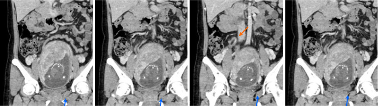
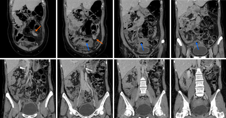
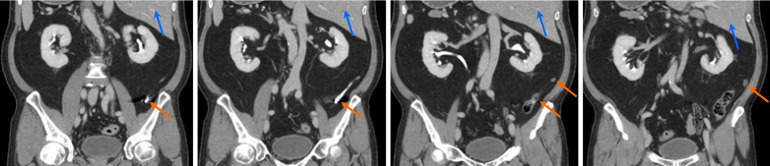
Results: Idiopathic pain was the most common diagnosis (104 cases, 45.2%), followed by appendicitis (46 cases, 20.0%) and diverticulitis (27 cases, 11.7%). Right lower quadrant pain predominantly revealed appendicitis (20.2%), whereas left lower quadrant pain frequently indicated diverticulitis (12.1%). Nonspecific pain cases have diverse etiologies. Rare conditions included situs inversus (one case) and intestinal malrotation (one case). Pregnancy-related diagnoses included acute appendicitis and uterine fibroid degeneration.
Conclusion: CT significantly aids in the diagnosis of lower abdominal pain, especially given limited access to ultrasonography and MRI during emergency hours in Japan. Awareness of the anatomical variations and pregnancy-related constraints is crucial. Diagnosis-specific protocols for CT based on pain location can optimize clinical management and resource utilization.

 求助内容:
求助内容: 应助结果提醒方式:


