Somaya Al Kiswani, Maysoon Nasser, Abdulla Alzibdeh, Elias Eq Lahham
{"title":"Enhancing back pain and sciatica diagnosis: Coronal short tau inversion recovery's role in routine lumbar magnetic resonance imaging protocols.","authors":"Somaya Al Kiswani, Maysoon Nasser, Abdulla Alzibdeh, Elias Eq Lahham","doi":"10.4329/wjr.v17.i6.107164","DOIUrl":null,"url":null,"abstract":"<p><strong>Background: </strong>Back pain and sciatica are common complaints that often require imaging for accurate diagnosis and management. Conventional lumbar magnetic resonance imaging (MRI) protocols typically include sagittal and axial T1 and T2 sequences; however, these may miss certain pathologies. The addition of coronal short tau inversion recovery (STIR) sequences offers the potential to enhance the detection of both spinal and extra-spinal abnormalities, thereby improving clinical decision-making and patient outcomes.</p><p><strong>Aim: </strong>To evaluate the impact of adding coronal STIR sequences to routine lumbar MRI in diagnosing back pain and sciatica.</p><p><strong>Methods: </strong>We prospectively analyzed data from patients aged 6 and older presenting with back pain or sciatica who underwent lumbar spine MRI at our institution. The standardized MRI protocol utilized included sagittal and axial T1 and T2 sequences, complemented by a coronal STIR sequence. Data on structural abnormalities were collected, reviewed, and analyzed using counts, percentages, and Fisher's exact test for categorical variables.</p><p><strong>Results: </strong>Our cohort comprised 274 patients (115 males, 159 females; mean age 44.91 years). Notably, 39 patients exhibited abnormalities across all sequences, while 72.63% showed normal findings on the coronal STIR sequence. Importantly, 30.29% of cases were diagnosed as normal without the coronal STIR, and 36 patients with normal T1 and T2 sequences presented abnormalities on the coronal STIR. The coronal STIR sequence successfully identified 26 spinal and 10 non-spinal pathologies, including 17 cases of sacroiliitis, with a significant association (<i>P</i> < 0.0001) between sacroiliitis diagnosis and abnormalities visible solely on this sequence.</p><p><strong>Conclusion: </strong>Integrating coronal STIR into routine lumbar MRI enhances detection of hidden spinal and extra-spinal pathologies, improves patient management, and offers a cost-effective, practical upgrade with significant diagnostic and clinical value.</p>","PeriodicalId":23819,"journal":{"name":"World journal of radiology","volume":"17 6","pages":"107164"},"PeriodicalIF":1.5000,"publicationDate":"2025-06-28","publicationTypes":"Journal Article","fieldsOfStudy":null,"isOpenAccess":false,"openAccessPdf":"https://www.ncbi.nlm.nih.gov/pmc/articles/PMC12210196/pdf/","citationCount":"0","resultStr":null,"platform":"Semanticscholar","paperid":null,"PeriodicalName":"World journal of radiology","FirstCategoryId":"1085","ListUrlMain":"https://doi.org/10.4329/wjr.v17.i6.107164","RegionNum":0,"RegionCategory":null,"ArticlePicture":[],"TitleCN":null,"AbstractTextCN":null,"PMCID":null,"EPubDate":"","PubModel":"","JCR":"Q3","JCRName":"RADIOLOGY, NUCLEAR MEDICINE & MEDICAL IMAGING","Score":null,"Total":0}
引用次数: 0
Abstract
Background: Back pain and sciatica are common complaints that often require imaging for accurate diagnosis and management. Conventional lumbar magnetic resonance imaging (MRI) protocols typically include sagittal and axial T1 and T2 sequences; however, these may miss certain pathologies. The addition of coronal short tau inversion recovery (STIR) sequences offers the potential to enhance the detection of both spinal and extra-spinal abnormalities, thereby improving clinical decision-making and patient outcomes.
Aim: To evaluate the impact of adding coronal STIR sequences to routine lumbar MRI in diagnosing back pain and sciatica.
Methods: We prospectively analyzed data from patients aged 6 and older presenting with back pain or sciatica who underwent lumbar spine MRI at our institution. The standardized MRI protocol utilized included sagittal and axial T1 and T2 sequences, complemented by a coronal STIR sequence. Data on structural abnormalities were collected, reviewed, and analyzed using counts, percentages, and Fisher's exact test for categorical variables.
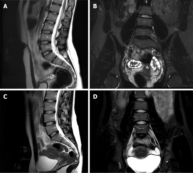
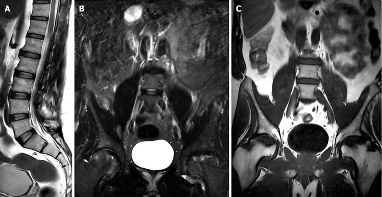
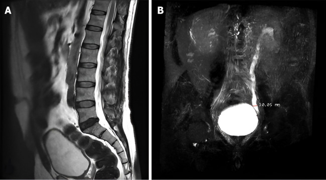
Results: Our cohort comprised 274 patients (115 males, 159 females; mean age 44.91 years). Notably, 39 patients exhibited abnormalities across all sequences, while 72.63% showed normal findings on the coronal STIR sequence. Importantly, 30.29% of cases were diagnosed as normal without the coronal STIR, and 36 patients with normal T1 and T2 sequences presented abnormalities on the coronal STIR. The coronal STIR sequence successfully identified 26 spinal and 10 non-spinal pathologies, including 17 cases of sacroiliitis, with a significant association (P < 0.0001) between sacroiliitis diagnosis and abnormalities visible solely on this sequence.
Conclusion: Integrating coronal STIR into routine lumbar MRI enhances detection of hidden spinal and extra-spinal pathologies, improves patient management, and offers a cost-effective, practical upgrade with significant diagnostic and clinical value.

 求助内容:
求助内容: 应助结果提醒方式:


